We perform tooth extraction and anesthesia treatment for patients who have difficulty in treatment at general dental clinics, such as heart disease, high blood pressure, diabetes, and taking medicine that makes it difficult for blood to stop. We work closely with local dental clinics, so please be sure to bring a letter of introduction. We do not provide general dental treatment such as treatment of cavities, coverings, dentures, bridges, dentures, periodontal disease, implants, etc.
Addressing Oral Surgery Diseases
We provide standard treatment at a high level for oral surgical diseases such as stomatognathic inflammation, jaw cysts, oral tumors, dental/oral and maxillofacial trauma, jaw deformities, temporomandibular joint diseases, salivary gland diseases, and oral mucosal diseases. I am trying to do it. All treatments for oral cancer and other oral malignant tumors are concentrated in Head and Neck Surgery and Head and Neck Surgery and Neck Surgery, we jointly participate in surgery and are in charge of jaw resection and jawbone reconstruction.
Initiatives for Minimally Invasive Treatment
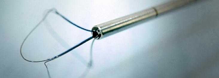
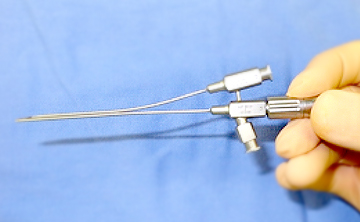
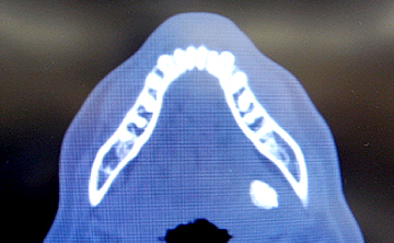
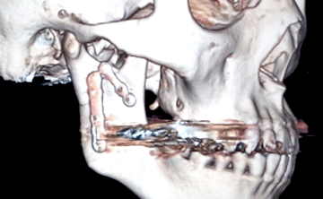
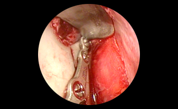
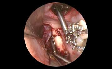
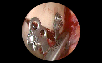
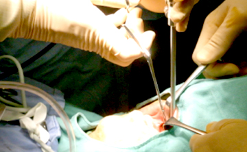
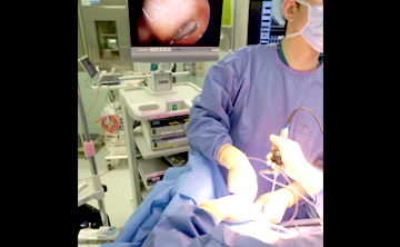
In addition, since around 2003, our department has been working on minimally invasive oral surgery using an endoscope. Endoscopic-assisted open reduction and fixation of mandibular process fractures, which involves reduction and fixation from the oral cavity under endoscopic support, is a facial skin incision It does not leave a scar because it fixes the fractured part without performing a procedure. In maxillomandibular fracture surgery, we aim to establish an appropriate occlusion at the time of surgery, and aim for treatment without postoperative intermaxillary fixation (a treatment that binds the upper and lower teeth with a wire to prevent the mouth from opening). Therefore, the patient can take it orally from the next day.
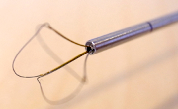
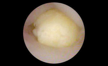
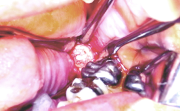
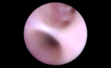
In the treatment of sialolithiasis, we are working on many sialolithectomy procedures using salivary gland endoscopy and sialolithectomy procedures through intraoral incision. In most cases, submandibular gland removal can be avoided, and about 50 cases are treated annually. Sialolithectomy is performed under inpatient general anesthesia, and the hospital stay is 3 nights and 4 days.
We have experienced many surgical corrective surgeries for jaw deformities.
We provide surgical orthodontic treatment for skeletal malocclusion (jaw deformity) that cannot be improved by orthodontic treatment alone, in collaboration with orthodontic specialists. More than 120 surgical cases are performed annually. In the upper jaw, Le Fort type I osteotomy is mainly performed. Regardless of the combination of surgeries, the hospital stay is about one week and you can eat with your mouth open from the day after the surgery. Autologous blood transfusion is not required. In addition, for severe jaw deformity, which is a congenital disease, we aim to improve not only the skeleton but also the soft tissue in cooperation with bone lengthening surgery for the jawbone and Plastic Surgery.
We have a 2-year Junior Resident system and a 3-year Senior Resident system.
Our department is certified as a designated training institution by the Japanese Society of Japanese Society of Oral and Maxillofacial Surgeons and the Japanese Society of Maxillofacial Implants. In addition, hospital dentists have established a training doctor system aiming for five years of oral Certified Surgeon, which is rare in Japan. Every year, two Junior Resident (two-year program) and one Senior Resident (three-year trainee program) are recruited from among excellent human resources nationwide. We also accept student internships.
Oral and Maxillofacial Surgery: High Standards of Care
<medical staff>
<Endoscopic surgery>
Medical record
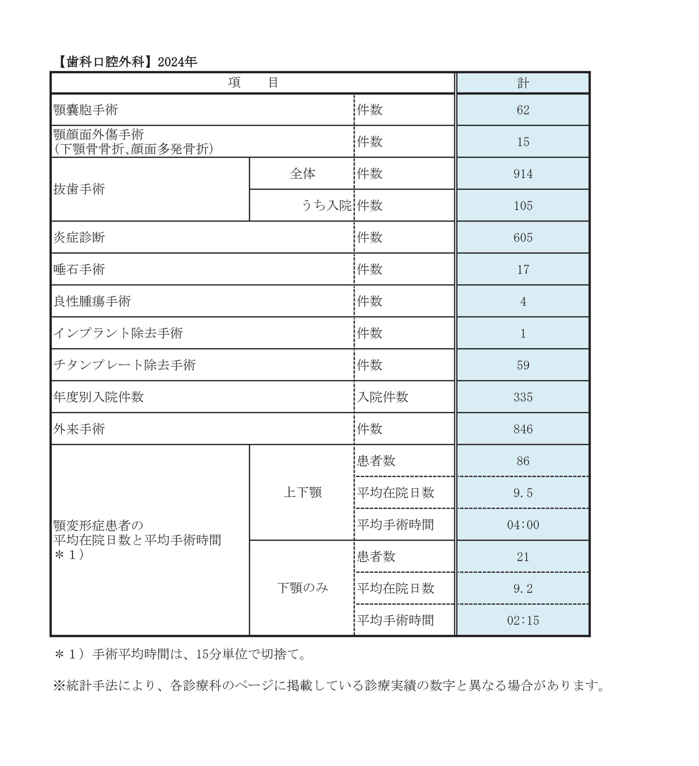
Dental and Oral Surgery 2021 Inpatient Surgery
alveolar bone surgery (sinus lift/bone graft) | 1 example |
---|---|
jaw cyst surgery | 44 cases |
jaw deformity surgery | 147 cases |
maxillofacial trauma surgery | 11 cases |
tooth extraction surgery | 38 cases |
salivary stone extraction | 25 cases |
benign tumor surgery | 4 cases |
implant surgery | 1 example |
titanium plate removal surgery | 67 cases |
Number of hospitalizations by year | 338 cases |
outpatient surgery | 563 cases |
Breakdown of Jaw Deformity Surgery
number of cases | |
maxillomandibular surgery | 121 cases |
---|---|
Mandibular surgery alone | 26 cases |
Departmental statistics
Main diseases/treatments
jaw deformity
A combination of orthodontic treatment and surgery to correct the jawbone can provide a correct bite.
Why should I have surgery?
Malocclusion refers to a problem with the way the teeth fit together, and malocclusion, which can be corrected by orthodontic treatment alone, is called orthodontic malocclusion. On the other hand, problems with the shape and positional relationship of the upper and lower jaws are called skeletal malocclusion or jaw deformity. In such cases, orthodontic treatment alone cannot cure the problem because of the problem in the skeleton, and correct occlusion can be obtained by combining orthodontic treatment with surgery to correct the positional relationship of the jawbone. increase. This is called surgical orthodontic treatment for jaw deformity.
Surgical orthodontic treatment is a treatment to improve the occlusion, so it is not a surgery with the main purpose of cosmetic surgery (for this reason, health insurance is applied). Therefore, if there is no malocclusion (simply square jaws, etc.), treatment is generally not possible. Cooperative treatment by an orthodontist and a dental or oral surgeon is always required.
Surgical orthodontic treatment involves moving the jawbone, which changes many things, including the face. For example, to improve the mouth, the lower jaw is retracted, so the inside of the mouth feels narrower. Surgery on the upper jaw will naturally change the shape of the nose and upper lip, and it may also cause nasal congestion and decreased nasal passage. Scars and scars remain where the scalpel was inserted, and adhesions occur where detachment occurs. Starting with changes in occlusion, numbness, twitching, various discomforts, changes in soft tissues such as facial movements, expressions and shapes, pronunciation, mastication, breathing, swallowing, temporomandibular joints, etc. is no exaggeration. Each person has a different way of feeling these things, and it is difficult for anyone other than yourself to understand them. Also, changes cannot be accurately predicted preoperatively, and there are no tests to evaluate and substantiate them postoperatively. Most people gradually adapt to these phenomena, and although they may feel discomfort and inconvenience for a while, they are pleased with the many improvements, including occlusion, and say that they are glad that they received treatment. The majority. However, there are some people (less than 1%) who cannot adapt to the change (even medical staff cannot predict it in advance). However, for subjective things such as numbness and other sensory changes and changes in appearance, there is no test/evaluation method to identify the cause, and there is no treatment that can be said to be absolutely effective, so once surgery is complete. cannot be returned to its original state. In addition, since minor aesthetic dissatisfaction is covered by insurance, reoperation cannot be performed to improve it. It is very important that you understand and agree to these things before you receive treatment. Not only those who have undergone surgery at our department, but there are many cases where the above consultations come after treatment at other medical institutions, but even if these events occur, there is still a desire to improve the occlusion. If you do not have orthodontic treatment, it is important that you decide on your own to choose only orthodontic treatment without surgical orthodontic treatment.
What is the flow of treatment?
Surgical orthodontic treatment consists of four major steps. Normally, the entire process takes about three years.
Stage 1 (preoperative orthodontic treatment)
Before surgery, we correct the alignment and inclination of the teeth and flatten the occlusal plane to create a stable occlusion after surgery. Preoperative orthodontic treatment takes an average of one and a half to two years.
There are people who wish to have surgery when preoperative correction is insufficient due to school or work reasons, but we do not recommend it. Surgical orthodontic treatment is covered by health insurance between our clinic and a licensed orthodontic clinic that meets the requirements. In Japan, insurance medical care is applied to medical care for which standard methods have been established. Surgery for cosmetic purposes, etc.).
At the first visit to our department, we will make an appointment for X-ray, 3D CT, and MRI of the temporomandibular joint.
Stage 2 (hospital surgery)
Once the completion of preoperative orthodontic treatment is in sight, the orthodontist will notify you to schedule the surgery. At that stage, we will discuss the surgery date at our hospital. Currently, there is a waiting period of about one year or more for inpatient surgery in our department. Please accept our apologies for not being able to accommodate your desired surgery schedule in many cases.
Approximately 3 months before hospitalization, a set of preoperative examinations (blood test, urine test, chest X-ray, electrocardiogram, maxillofacial CT, etc.) for general anesthesia will be performed, and the details of the surgical plan and surgical risks will be explained again. We will also provide you with a consent form. You will be admitted to the hospital the day before the operation and will be hospitalized for about 10 days.
In addition, since the surgery will be scheduled for more than a year ahead, there is a possibility that the outpatient doctor and the surgery doctor will change.
Stage 3 (postoperative orthodontic treatment)
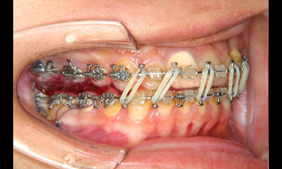
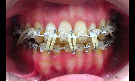
After the surgery, you will undergo rehabilitation with a mini rubber band for correction (described later) for about 3 months. You can also have your orthodontist make minor adjustments to the alignment of your teeth. If a titanium plate for osteosynthesis is used in surgery, plate removal surgery is performed 7-12 months after surgery.
Stage 4 (retention)
The orthodontic wire is removed, and the retainer is used only during the day or night to stabilize the alignment of the teeth, and the treatment is completed.
What is the target disease (jaw deformity)?
When the upper and lower jawbone such as socket (mandibular protrusion) and protruding teeth (maxillary protrusion) are too large or too small (maxillary recession due to cleft lip and palate or micromandible). There are various cases, such as when the face or jaw is crooked (facial asymmetry, mandibular asymmetry) or when the upper and lower front teeth do not fit together (open bite).
What is the surgical method?
Surgery is performed under general anesthesia. There are several surgical methods for various jaw deformities, but in principle, the surgery is performed from the inside of the mouth and does not cut the facial skin. However, this is not the case in emergencies, such as when a large amount of bleeding requires hemostasis, or when there is a possibility of airway obstruction after surgery, and skin incision or tracheotomy may be required. Cases of death or vegetative state due to airway obstruction due to hemorrhage etc. have been reported).
The surgical methods listed below are combined to accommodate various deformities.
First, I will explain the surgical method for the upper jaw.
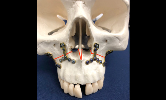
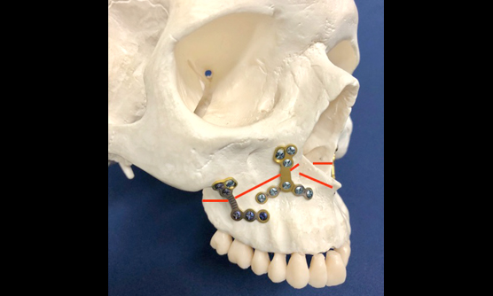
Le Fort type I osteotomy
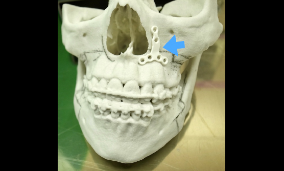
This is a method for moving the entire upper jaw. Make an incision in the gum inside the upper lip and cut the bone horizontally from the side of the nose. Since the entire upper jaw can be moved with the teeth attached, after moving it to the correct position, it is firmly fixed with a plate and screws for osteosynthesis. At our clinic, we mainly use bioabsorbable plates for the upper jaw.
Maxillary anterior alveolar osteotomy
This method is used when there is a problem only in the front teeth of the upper jaw. In the surgery, the left and right first premolars (fourth teeth) are extracted, the alveolar bone is removed, the six front teeth from the left and right canines to the canines are separated, the teeth are extracted, and the bone is removed to create a space. to the After moving to the correct position, the bone fragments are fixed with plates and screws, and further fixed with a custom-made plastic palatal base from the back side of the teeth. The palatal floor is removed on an outpatient basis after one month.
Next, I will explain the surgical method for the mandible.
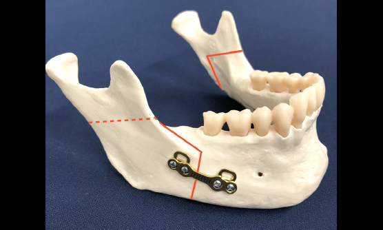
Mandibular branch sagittal segmentation
This is the most standard method of moving the entire lower jaw. An incision is made in the gums and buccal mucosa around the wisdom tooth, and the bone is divided into inner and outer parts. After osteotomy on both the left and right sides, only the bone with the teeth is moved to the correct occlusal position without moving the bone with the outer temporomandibular joint. The bone is then fixed with plates and screws.
Intermaxillary fixation (a treatment to bind the upper and lower teeth with thin wire) will not be performed on the day of surgery, and rehabilitation using orthodontic mini rubber bands will begin the day after surgery. Wisdom teeth must be removed six months prior to this surgery.
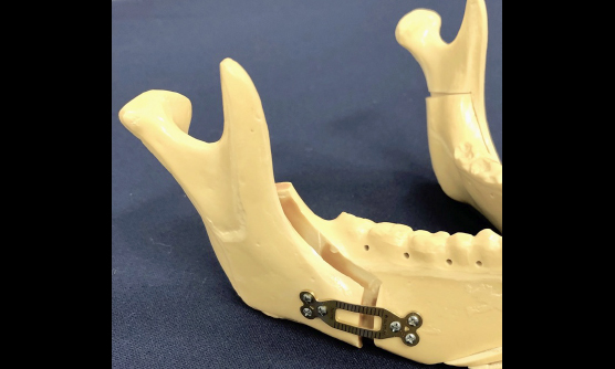
In our department, we use a titanium plate and an absorbable plate depending on the case, because a large force of biting is applied during surgery on the lower jaw. Titanium plates have approximately five times the physical strength of absorbent plates. In practice, we determine during surgery how much load will be applied, and then select the plate. If a titanium plate is used, a separate removal surgery will be performed.
In addition, although there is no need to remove the absorbable plate, in rare cases, postoperative foreign body reaction or infection may occur, requiring additional surgery or treatment.
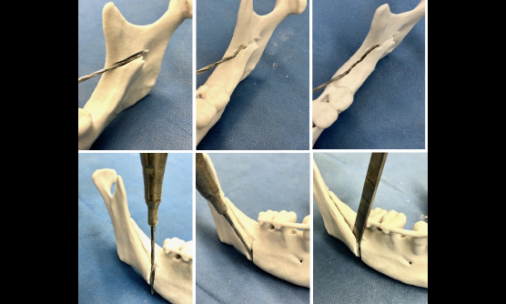
Mandibular ramus vertical osteotomy
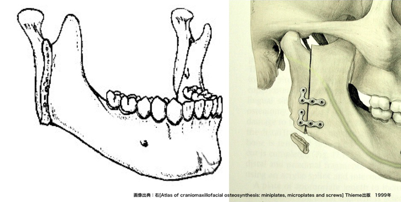
It is also a way to move the entire lower jaw. After all, an incision is made in the gums and buccal mucosa around the wisdom teeth, and the bone in front of the temporomandibular joint is cut vertically in a straight line from top to bottom, and the lower jaw is separated into the left and right joints and the part where the teeth are planted. After that, the bones in which the teeth are planted are moved to the correct occlusal position. In most cases, this method does not fix the separated bones with a plate or the like, but the bones fuse without problems. Mandibular sagittal segmentation is the standard surgery for the mandible not only in Japan but also in the world. Although this vertical osteotomy is not very popular, it is a useful method when the mandibular bone is thin and easily broken. From the day of the surgery to the next morning, you will rest with your mouth closed using a mini orthodontic rubber band. Vertical osteotomy frees the bones in the joint area, so movement of the jaw does not affect the temporomandibular joint. may be used.
Mandibular anterior alveolar osteotomy
The mandibular anterior alveolar osteotomy is similar to the maxillary anterior alveolar osteotomy.
Menoplasty
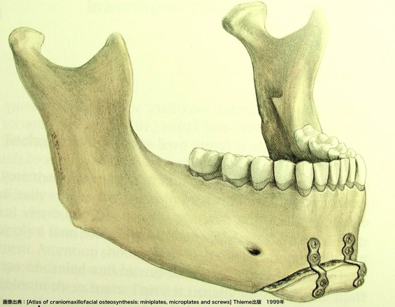
An incision is made in the gums inside the lower lip and the operation is performed. Correct the position and shape of the tip of the mandible called the chin. This surgery is not related to occlusion, but is sometimes done to balance the facial skeleton after jaw movement with other surgical methods. Unlike cosmetic surgery that inserts artificial objects, we move and modify your own bones and fix them with plates and screws (titanium or bioabsorbable).
Menoplasty is performed at the time of plate removal surgery if the attending physician deems it necessary after evaluating the facial appearance after improving the occlusion by moving the upper and lower jaws. By performing chin formation after the initial surgery, it is possible to make fine adjustments while looking at the overall balance.
However, since there is a limit to chin formation, it is unpredictable whether you will have the desired facial appearance after surgery. In addition, since the operative field is close to the neural foramen (the exit of the nerve), there is a high risk of postoperative discomfort and perceptual abnormalities.
Jaw lengthening surgery
In the treatment of micromandibular disease with a small jawbone, conventionally, the mandibular ramus sagittal division was used to extend the mandible forward. However, with this method, the muscles and skin that attach to the jawbone, and the nerves and blood vessels that run through the jawbone, are forcibly stretched, limiting forward movement (up to about 15 mm). In addition, even after the surgery, some of the jawbone, which had been moved, would return to its original position due to the pull-back force of the skin and muscles. A Le Fort I osteotomy has traditionally been performed to move the small upper jaw forward in patients with cleft lip and palate, but scarring from previous surgeries often prevents sufficient movement. There was nothing. In jawbone lengthening, an osteotomy line is made on the part of the bone to be lengthened, and an implanted bone lengthener is attached there. After the surgery, the lengthener is activated and the lengthening is started at a very slow speed of about 1mm per day. The gaps between the bones will open day by day, but eventually new bone will regenerate there. Moreover, not only bones but also tissues such as nerves and blood vessels are regenerated. By using this method, it has become possible to treat severe jaw deformities such as Treacher Collins syndrome, which were previously impossible to treat.
Surgery combines these methods to address various deformities.
plate removal surgery
We often receive the question, "Will my nose be deformed after maxillary surgery?" In conclusion, it can be said that subtle changes in the shape of the nose are unavoidable as long as the maxillary surgery is performed. Changes in the shape of the nose are thought to refer to widening of the wings of the nose (parts of the nostrils), upward pointing of the tip of the nose, and changes in the shape of the nostrils. After the surgery, the upper lip and the cheeks around the nose are swollen to about 1.5-2 times the thickness before the surgery, so you should wait until 4-6 months after the surgery for them to completely subside. . However, there are two main reasons why the shape of the nose looks different after surgery. The first is that the movement of the upper jaw changes the morphology of the mouth, especially the angle formed by the nose and upper lip, which changes the shape and appearance. The nose is a tent of cartilage that sits on top of a gaping pear-shaped hole in the middle of the upper jaw. If the upper jaw, which is the ground, moves, the inclination and shape of the tent on top of it will naturally change. This change becomes more noticeable as the maxillary displacement increases.
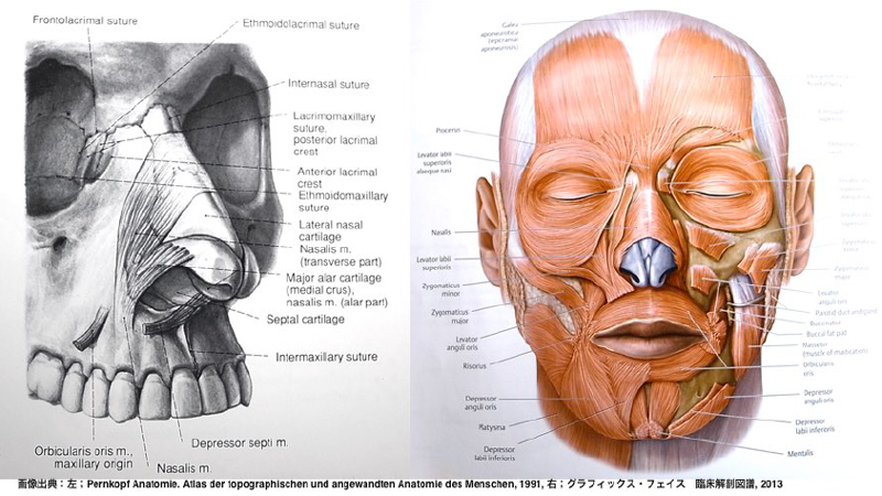
The second reason is that the tightness of the entire nose is loosened. Even if the ground doesn't move much, if the ropes that hold the tent loose, the shape of the tent itself will change. When you smile, your nostrils open and the corners of your mouth also move upwards. This is because facial muscles (also called facial muscles) move. Facial muscles are muscles that originate from the facial bones and attach to the facial skin. There are many facial muscles on the face, especially around the nose and corners of the mouth. Of course, the nose also has a facial muscle called the nasal bridge, and around the nasal alar, facial muscles such as the levator nasalis superior muscle and the levator labialis superior muscle attach from the maxillary bone to the skin of the nose and nasal alar. A facial expression is created by pulling the skin using the attachment to the bone as an anchor. During maxillary surgery, these attachments of facial muscles must be removed from the bone in order to cut the bone. In order to return the stripped facial muscles to their original positions as much as possible after fixing the plate, it is necessary to pull the stripped left and right muscles with a thread. In our department, both the left and right are pulled and fixed with two threads. It also reattaches the cartilage in the middle of the nose, called the nasal septum, to the maxillary bone. In addition, a suture is also performed to pull the back of the upper lip at the midline. These three traction sutures restore tension and tighten the nose tent. However, even with such meticulous attention to detail, there are still a few percent of patients who are concerned about the shape of their nose after surgery.
Although the majority of people do not notice it, changes and deformations of the nose will inevitably occur after maxillary surgery, and in some cases, nasal congestion and decreased nasal passage may occur due to changes in the volume of the nose. Please understand.
What are the risks of surgery?
Next, the risks of surgery are explained.
1. Risk of excessive bleeding during surgery
In fact, there is little to worry about, but in the unlikely event that a large amount of bleeding occurs, hemostasis is given priority, and the operation may be interrupted and postponed to a later date.
2. Possibility of revision surgery
In the case of our department, the probability is around 1.5%. Hemostasis treatment for abnormal postoperative bleeding, removal of stray foreign objects such as orthodontic devices, dislocation of the temporomandibular joint, occlusion defects not seen during surgery, and bone fragment defects that cannot be identified without postoperative imaging. For example, if repositioning and fixation is required, etc. Most of these occur during hospitalization, but in rare cases postoperative infection after discharge, abnormal fractures, failure of bone union, and malocclusion due to broken plates may require reoperation.
3. Some parts of the body, such as the lips, the skin and mucous membranes of the cheeks, the gums and the tongue, always have numbness. Depending on the shape of the bone, the course of the nerves, and the difference in the surgical method, the place where the numbness is strong varies from person to person.
Usually, most of the symptoms will be relieved in about six months to a year, but some people may experience paresthesia or paralysis as aftereffects. Recovery tends to be delayed with age, such as in the 40s and 50s.
There is a small chance that facial nerve palsy (impaired movement of the muscles that produce facial expressions) will occur and have sequelae.
Four. temporomandibular joint symptoms
After surgery, temporomandibular joint symptoms such as pain, noise, and trismus may appear or worsen. Unfortunately, complete surgical control of arthritis is not possible. If symptoms develop, we will take the best measures according to the condition of the patient.
Five. tooth damage
If the osteotomy is close to the tooth, postoperative pulp necrosis, periodontitis, root resorption, and tooth fracture may occur. If these symptoms are confirmed, treatment such as tooth restoration treatment, root canal treatment, or tooth extraction may be required.
6. Fear of postoperative infection
If you have caries or infected foci on your teeth, or if you keep your mouth dirty, your teeth may cause wound infections. Wisdom teeth in the lower jaw must be extracted 6 months before surgery. In addition, since bacteria exist in saliva in the first place, intraoral surgery may inevitably cause postoperative bacterial infection. If infection does occur, surgery to clean the wound and remove fixed plates may be required.
7. Swelling of the face, sagging around the cheeks and chin, and subtle changes in the shape of the nose after surgery on the upper jaw are inevitable. Since the upper jaw moves, some changes in appearance and facial expressions, including the nose, cannot be avoided. This surgery is necessary to improve the occlusion.
Depending on the amount and direction of movement of the jaw, the swelling after the surgery will be reduced to a level that strangers will not notice in about a month. The swelling that you or someone close to you notices generally lasts for about half a year after the surgery.
8. Inability to eat and talk for about a month
About a month after the surgery, you can almost return to your original diet except for things like tearing off hard meat. Before you leave the hospital, it is often possible to talk on the phone.
9. Postoperative relapse
Soft tissues such as muscles are attached to the displaced bones, so the surgery creates tension in the soft tissues and forces the bones to return to their original positions. This often causes setbacks. The amount of relapse differs depending on the case, and it can be dealt with by postoperative orthodontic treatment, but in some cases it cannot be dealt with.
In rare cases, progressive morphological changes and resorptions occur in the head of the temporomandibular joint (progressive mandibular condyle resorption). may also be required. The cause has not been elucidated, but it is reported that it is highly likely to occur in patients with a small mandibular condyle or after surgery for mandibular recession or open bite.
In order to reduce these risks as much as possible, rehabilitation of rubber band hanging (described later) is important.
Ten. others
Surgical orthodontic treatment changes various things including the face, although it is not a cosmetic surgery as it moves the jawbone to improve the occlusion. Retracting the lower jaw to improve the socket will make the mouth feel narrower. After maxillary surgery, the shape of the nose and upper lip will naturally change, and nasal congestion and decreased nasal passage may occur. It is no exaggeration to say that everything changes, starting with occlusion, numbness, twitching, various discomforts, facial movements, facial expressions and shapes, pronunciation, chewing, breathing, swallowing, and the condition of the temporomandibular joints. Each person has a different way of feeling these things, and it is difficult for others other than yourself to understand them. Also, changes cannot be accurately predicted preoperatively, and there is no way to evaluate or demonstrate them postoperatively. Most people adapt to these events gradually, but some people (less than 1%) cannot adapt to the changes (even medical professionals cannot predict them in advance). However, subjective things such as numbness and other sensory changes, changes in appearance, and dissatisfaction are completely eliminated once surgery is performed because there is no test to identify the cause or treatment that can be said to be absolutely effective. It is impossible to return it to its original state. In addition, since minor aesthetic dissatisfaction is covered by insurance, reoperation cannot be performed to improve it. Please understand and acknowledge these things in advance before receiving treatment.
We will explain the above 10 items at the first visit.
All surgeries are performed under general anesthesia by a specialist Anesthesiology. In the past, for maxillary and mandibular surgeries, which are likely to cause increased bleeding, 800ml of autologous blood was collected before the surgery to prepare, and after surgery, the patient stayed in the intensive care unit (ICU) for one night with an endotracheal intubation. Currently, we do not perform autologous blood collection or postoperative endotracheal intubation tube placement, but for safety reasons, we may conduct follow-up observations in the high care unit (HCU) adjacent to the operating room on the night of the surgery. Similarly, there is no infusion of liquid food by indwelling nasogastric tube through the nose, and the patient will be given oral intake from the morning after surgery. Since 2003, we have introduced a clinical path that standardizes treatment methods, and we are working to ensure that patients can spend their time in the hospital with peace of mind while checking the course of treatment themselves.
How is your recuperation life in the hospital?
You will be able to rest in bed for a few hours immediately after surgery, but after that you will be able to get up and walk gradually. Many people are worried about pain immediately after surgery, but in fact, there are not many people who are troubled by pain. In most cases, intravenous drips and suppositories can be used immediately after surgery, and general oral pain relievers can provide sufficient pain relief from the next day. After discharge from the hospital, some patients do not need pain relievers.
Rather than the pain, many people said that nausea after waking up from anesthesia, stuffy nose after surgery, and nosebleeds dripping into the throat were the most painful. It is said that nausea after anesthesia tends to occur in people who are prone to car sickness, but we have prepared several types of anti-nausea drips, so we will respond appropriately while monitoring the progress. In addition, since the wound on the lower jaw is closed, a tube called a continuous suction drain is placed to suck out the blood that has accumulated in the wound. But inevitably drips down my nose and into my throat. If you swallow this blood, blood clots will gradually form in your stomach and cause further nausea, so use the aspirator prepared in advance by your bed and suck it out from your mouth or nose. is important. When it comes to nausea, there is a clear distinction between those who experience nausea and those who do not.
Nasal congestion is caused by the effects of the anesthesia nasotracheal tube and the swelling of the surgery itself. When you are admitted to the hospital, we will prescribe nasal drops, so please use them as needed. If you have a chronic nasal congestion such as allergic rhinitis, be sure to consult an otolaryngologist before hospitalization and receive prescriptions such as medicine. Also, if you have had surgery on your upper jaw, please do not blow your nose strongly while you are in the hospital. If you blow your nose strongly, air will enter the cheeks and under the eyes from the osteotomy, resulting in strong swelling called emphysema. Please refrain from blowing your nose strongly for about 2 weeks.
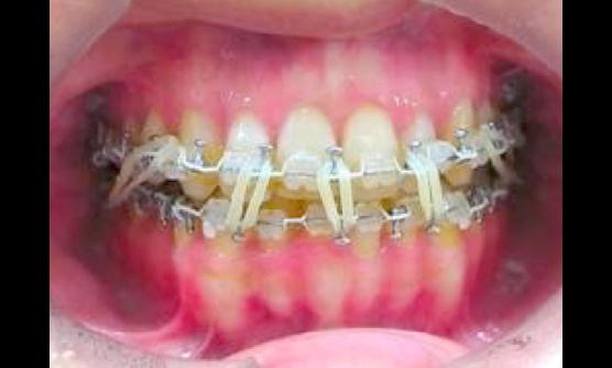
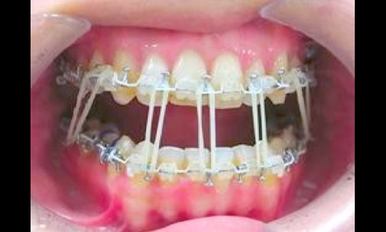
Even if skeletal correction is performed by surgery, the muscles around the jaw will not work for a while after surgery, so you will not be able to bite to the right position on your own and will be in a half-open state. Until the muscles get used to it, it is necessary to rest with the mouth closed while guiding the upper and lower jaws to the correct occlusal position by hanging the orthodontic mini rubber band on the hook attached to the orthodontic wire. From the next day onwards, your doctor will instruct you on how to put on the rubber band, so please remove the rubber band yourself when you eat, brush your teeth after eating, and put it back on yourself. At first, you may find it difficult to attach the rubber band, but if you can do it well by the time you leave the hospital, there will be no problem. As for the diet, liquid diet is started on the morning after surgery (no tube feeding through the nose), and from the second day onwards, it is increased to 3-minute porridge, and when it becomes possible to eat it, it is increased to 5-minute porridge. In addition, an anti-suppuration drip is available until the day after surgery. You can shower and wash your hair on the second day after surgery.
What will life be like after being discharged from the hospital?
Discharge is usually 8 days after surgery. Pain relief after discharge from the hospital is often not necessary, and it is possible to gradually return to normal food (approximately one month). In addition, the mouth will gradually open, and the power to chew will also come out. After you leave the hospital, first see an orthodontist. Follow your orthodontist's instructions on how to wear the rubber band. One month after the surgery, you can open your mouth to some extent even with the rubber on.
Rehabilitation of rubber band hanging is required for about 3 months. This rehabilitation is extremely important. Even if the surgery goes well doctor you may still have problems with your occlusion if you neglect to attach the rubber band. It's the same as needing to put a cast on and rest after having surgery for a broken arm or leg. In addition, you will have regular outpatient visits at our hospital, and six months after the operation, X-rays and CT will be taken to check the position and healing of the bones.
After discharge, it depends on the progress, but in most cases you can continue your daily life such as school and work, except for eating, although the swelling still remains.
Can I use health insurance?
Jaw deformity is a disease that causes an abnormal bite, so if you are diagnosed with "jaw deformity" at a licensed orthodontist's office and if surgery is deemed necessary by an oral surgeon, correction will be covered by health insurance. Treatment and surgery are available. Patients who are receiving orthodontic treatment at their own expense will also be treated at their own expense for inpatient surgery. The cost of hospitalization and surgery depends on the type and number of surgeries and the number of days of hospitalization, but if covered by insurance, you will have to pay 30% of the total medical expenses yourself, and will be reimbursed at a later date as it is covered by high-cost medical care. On the other hand, if it is applicable to self-pay medical treatment, the cost will be about 1.3 to 1.6 million yen for surgery on the lower jaw only, and about 2.8 to 3 million yen for surgery on both the upper and lower jaws. Also, please note that all outpatient consultation fees, blood tests, imaging tests, and medicines will be covered at your own expense. In order for orthodontic treatment to be covered by insurance, the orthodontist in charge must be designated as covered by insurance (see the note below), so please contact doctor for details.
Since we do not have an orthodontist in our department, cooperation with a nearby orthodontic clinic is essential.
Supplementary note: Designated institution capable of stomatognathic function diagnosis calculation
- Regarding the medical treatment prescribed in Article 36, Item 1 and Item 2 of the Ordinance for Enforcement of the Act on Support and Support for Persons with Disabilities (Ministry of Health, ministry of Health, Labor and Welfare notation No. 19 of 2006), the Act on Support and Support for Persons with Disabilities (Act No. Item 123) Medical institutions (limited to those in charge of orthodontic treatment) designated by the prefectural governor as stipulated in Article 54, paragraph 2.
- Having sufficient dedicated facilities for the treatment concerned.
- A system of cooperation with another insurance medical institution that is in charge of surgery such as jaw transection should be in place for the treatment concerned.
sialolithiasis
Sialolithiasis, which occurs in the major salivary glands such as the submandibular gland, is a disease in which salivary stones block the ducts, causing repeated swelling and pain during eating. It occurs predominantly in the submandibular gland, and more often occurs near the exit (transitional area) from the body of the submandibular gland to the main duct. The size varies from about 1 mm to over 1 cm. Intraductal salivary stones running in the shallow part of the floor of the mouth are mainly removed by intraoral incision, and for deep parts, transitional parts, and salivary gland bodies, submandibular gland extirpation through skin incision under the mandible is usually selected. increase. The salivary gland duct has a diameter of 0.5 mm at the exit to the oral cavity, and a diameter of about 1.5 mm at the main duct inside.
In salivary gland endoscopic sialolithectomy, an endoscope with a diameter of 1.6 mm or 2.5 mm is inserted while enlarging the opening of the salivary gland duct in the oral cavity, and the salivary stone grasping forceps are extended from inside the endoscope to remove the sialolith. It's a way to grab the
Indications for sialolithiasis
When the sialolith is less than 5 mm in diameter and is present in the duct anterior to the glandular body.
Large salivary stones other than those mentioned above or those present inside the body of the salivary glands cannot be removed using an endoscope alone. Extract it by palpation. Most of the salivary stones that cannot be removed with an endoscope alone can be removed by intraoral incision, avoiding removal of the submandibular gland. The hospital stay is 3-4 days.
Endoscopy-assisted therapy
Traditionally, most fractures of the mandibular process were non-invasive treatment with only intermaxillary fixation. Although this method facilitates reduction of the fracture, it leaves scars on the face and may cause facial paralysis. Endoscopically assisted surgery is a method of reducing and fixing the temporomandibular joint process through an intraoral incision. This method was developed and put into practical use in Germany, but it was treated by making a small incision in the skin in front of the ear and drilling a small hole through which the instrument penetrated. In our department, we are currently practicing complete intraoral surgery that does not involve skin incision due to our own ingenuity.
Surgery is indicated for the base of the temporomandibular joint process or the lower neck, but not for the upper neck or the head of the joint.
In bone lengthening surgery for congenital hypoplasia of the jawbone, unlike normal acquired diseases, the surgical field is narrow and morphologically difficult. It is now possible to perform osteotomy, bone extension device attachment, and osteosynthesis plate attachment with the aid of an endoscope, enabling safer and more reliable surgery.
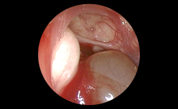
In the removal of odontogenic tumors in the mandible and maxilla, the use of an endoscope makes it possible to see parts that cannot be seen with the naked eye and expands the local field of view to detect tumors. There is no residue left behind, and the recurrence rate decreases. It also reduces the risk of nerve damage.
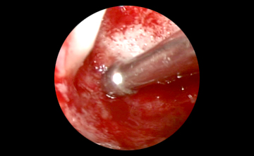
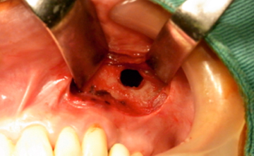
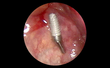
A 1-2 cm incision is made in the gingiva from the inside of the mouth, and a small hole with a diameter of about 5 mm is made in the wall of the maxillary sinus to remove foreign objects such as teeth and implants that have entered the maxillary sinus.
If you use an endoscope, you can remove it from a distance by opening a small window, so it does not interfere with subsequent dental or implant treatment.
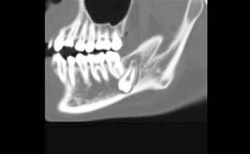
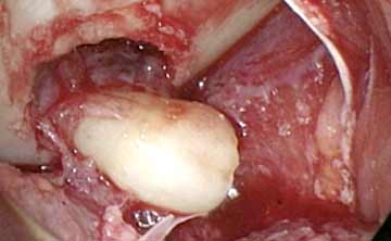
Endoscopy is also applied to the extraction of impacted wisdom teeth very deep in the lower jaw. Normally, it is a place where you can never see directly, but the tooth is extracted without cutting the skin while looking at the gingiva inside the back tooth with an endoscope.
We are currently not accepting new implant treatments.
clinical research
Notice regarding implementation of clinical research for patients undergoing dental and oral surgery
At present, we are conducting the following clinical research in the Department of Oral and Maxillofacial Surgery.
In this research, we will use the data (information) obtained from the patient's daily clinical practice.
If you object to the use of your data for this research, you can stop using the information or providing it to other research institutions at any time. If you would like to know more about the plan or content of the research, object to the use of your data in this research, or have any other questions, please contact us at the contact information below. .
Research subject name | Person in charge of our hospital | Explanatory text (PDF) |
Three-dimensional evaluation of maxillary movement and soft tissue changes in the nasolabial region following Le Fort I osteotomy | Shinsuke Yamamoto | |
---|---|---|
Indications and limitations of CAD/CAM splints in Le Fort I osteotomy | Shinsuke Yamamoto | |
Postoperative stability of mandibular width in midline mandibular fractures accompanied by bilateral condylar fractures | Keigo Maeda | |
Three-dimensional evaluation of postoperative stability of maxillary osteoplasty using u-HA/PLLA | Shinsuke Yamamoto | |
Usefulness of granisetron and ondansetron for prevention of nausea and vomiting after orthognathic surgery | Keigo Maeda |
Contact information at our hospital | Naoki Taniike Director Dentistry and Oral Surgery Kobe City Medical Center General Hospital 〒650-0046 4-6 Minatojima Nakamachi, Chuo Ward Kobe City TEL: 078-302-4321, FAX: 078-302-2487 |
---|