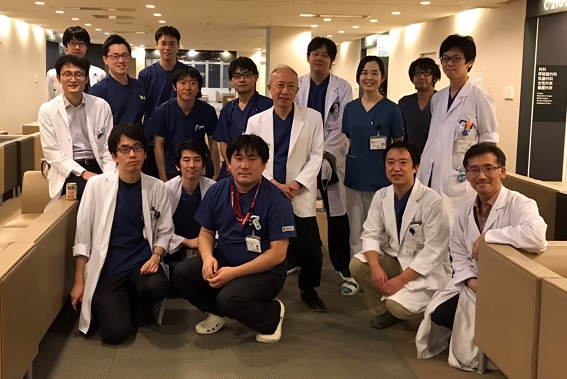
Based on the mission of a regional medical support hospital, we provide 24 hours a day, 365 days a year support for acute stages of all respiratory diseases, especially severe and intractable cases, rare diseases, etc. This is my motto.

Kobe City Medical Center General Hospital
Acting Director Respiratory Medicine
Ryo Tachikawa

Based on the mission of a regional medical support hospital, we provide 24 hours a day, 365 days a year support for acute stages of all respiratory diseases, especially severe and intractable cases, rare diseases, etc. It's my motto. The content of doctor care is based on the development of the latest diagnostic treatment based on evidence, and we always keep an awareness of problems and participate in many clinical trials and clinical trials to solve them. We strive to be a place where we can maintain a high level of motivation. We will continue to provide safe and advanced medical care so that we can be evaluated by citizens as the most reliable hospital for respiratory diseases.

Research by doctor Keisuke Tomii and doctor Kazuma Nagata of the Department of Respiratory Medicine at our hospital demonstrated the efficacy and safety of home high-flow therapy, a new treatment for COPD patients, and led to its insurance coverage.

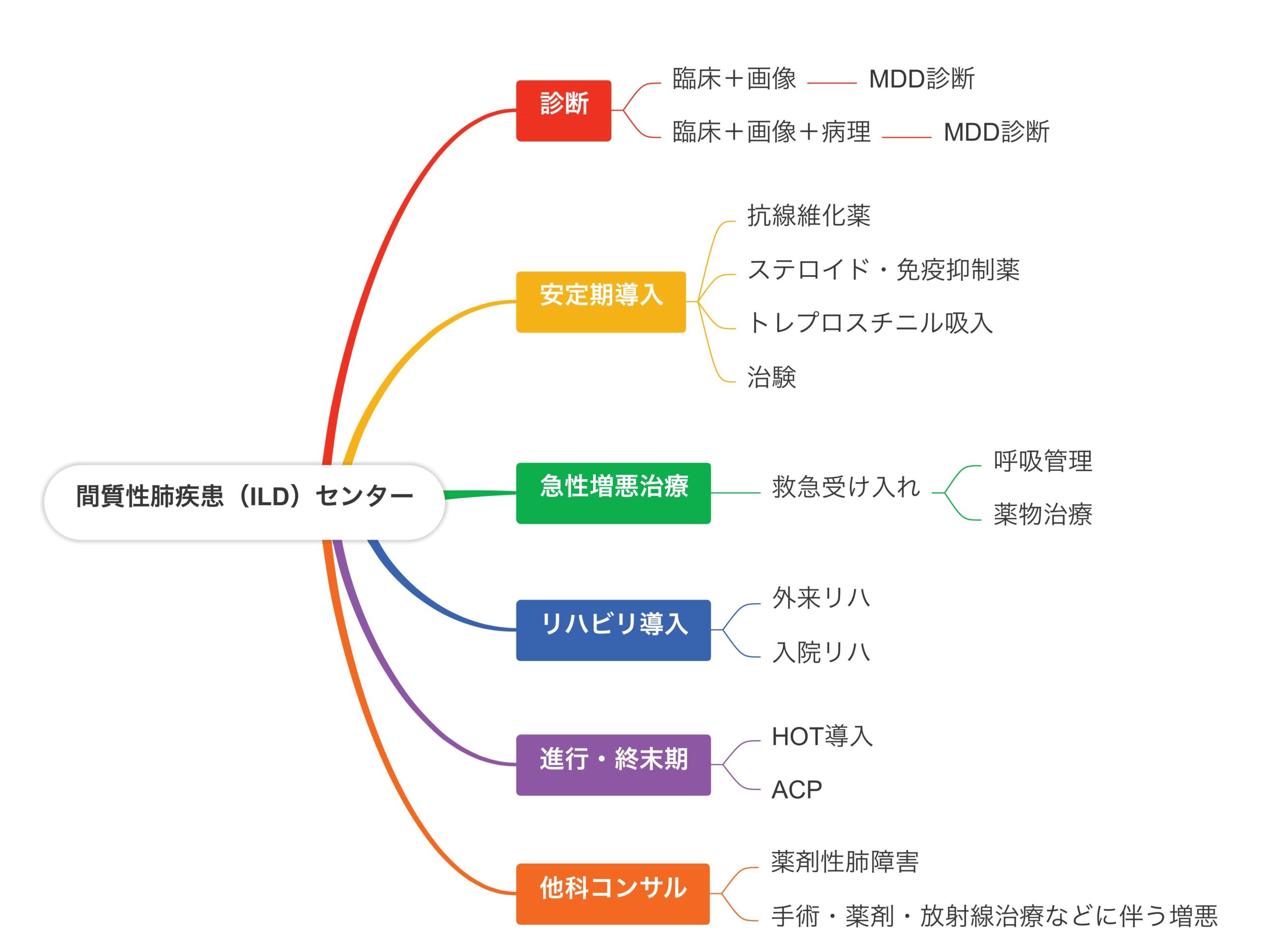
Regarding the opening of the Interstitial Lung Disease Center (ILD Center) (from April 2024)

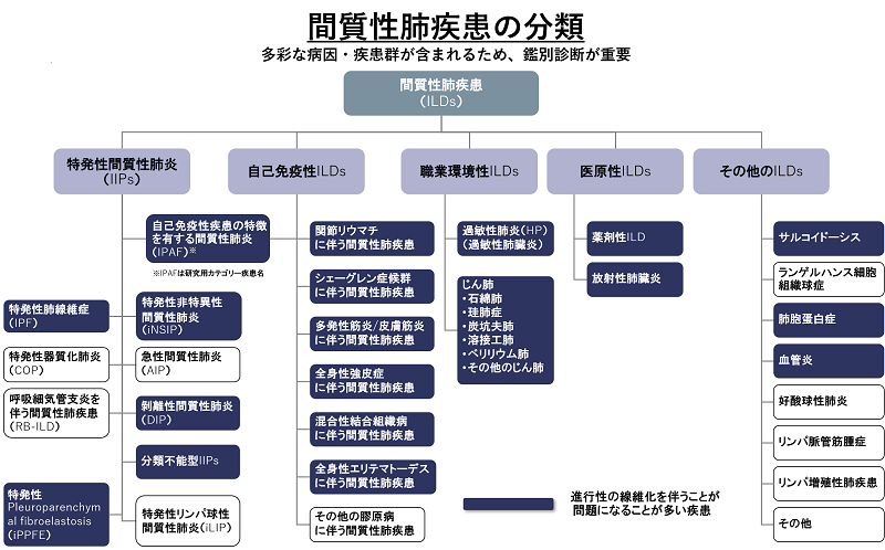
Interstitial lung diseases range from idiopathic interstitial pneumonia, which is an incurable disease, to those associated with collagen diseases, those associated with dust, smoking, and various antigen inhalations, those caused by drugs, and those associated with infectious diseases such as coronavirus. It may be a complication with other diseases or treatments, and its prognosis varies widely, including cases where it is cured, cases where fibrosis progresses and leads to respiratory failure, and cases where a sudden and fatal acute exacerbation occurs. Diagnosis requires specialized tests and consultation conducted at a facility experienced in medical treatment, and treatment requires specialized knowledge of various drugs, rehabilitation, respiratory management, etc., and the involvement of multiple professionals. In the case of progressive disease, lifelong support, education, and palliative care are also essential.
At our hospital, we have been involved in the medical treatment of some of the most prominent patients in the Kansai region as part of the field Respiratory Medicine, but this time we will further raise awareness of ILD and provide appropriate treatment to even more patients. We have set up a specialized center to provide care. Patients who are undiagnosed and have difficulty responding due to unclear images, patients whose respiratory failure progresses rapidly, patients whose ADL gradually declines and who have difficulty responding, patients who develop symptoms during treatment for cancer or collagen disease. We would like to support all kinds of ILD patients, both inside and outside the hospital.

Main target diseases and flow of medical treatment

About lung cancer

Lung cancer is a general term for cancer that develops in the lungs. Lung cancer occurs when normal cells in the trachea, bronchi, and alveoli lose their original functions and proliferate in an uncontrolled manner. Moreover, cancer cells do not stay in the local area, but spread to multiple organs along the flow of blood and lymph, and proliferate similarly. This is called transfer. Lung cancer is a highly malignant disease with a poor prognosis because of its rapid proliferation and metastatic potential.
The number of deaths from lung cancer in Japan has consistently increased for both men and women since the 1960s. In 1993, the death rate from malignant neoplasm by category surpassed gastric cancer to become the number one mortality rate for men, and third for women, and continues to increase. I'm here.

symptoms

Various symptoms appear depending on the size and location. Symptoms such as coughing and bloody sputum may be present, but in many cases there are no symptoms even if they are quite large.
Other common symptoms include chest and back pain, wheezing (wheezing), shortness of breath, hoarseness, and swelling of the face and neck. Common cancer symptoms include fatigue, loss of appetite, weight loss, and persistent fever.

Diagnosis of lung cancer
①Bronchoscopy

In order to obtain an accurate histopathological diagnosis of lung cancer, ultra-thin bronchoscopy, intrabronchial ultrasonography, and cryo biopsy are also performed for diagnosis.

(2) Ultrasound-guided biopsy

A biopsy is performed percutaneously under ultrasound guidance. It is performed for lesions that are close to the subcutaneous area.

③CT-guided biopsy

While taking a CT scan, the lesion is confirmed and a biopsy is performed percutaneously. Bronchial examination is performed for lesions that are difficult to reach.

④Thoracoscopic lung biopsy

In cooperation with our hospital's Respiratory Sugery, it is performed when it is difficult to make a diagnosis with the above tests.
In addition to the histological examinations mentioned above, diagnostic imaging such as X-ray examination, CT scan, MRI, bone scintigraphy, and PET/CT will be performed to diagnose the stage (progression of cancer).

Classification by tissue type

It is classified into four histological types: squamous cell carcinoma, adenocarcinoma, large cell carcinoma, and neuroendocrine carcinoma. In determining the treatment policy, it is often classified into two types: non-small cell lung cancer (squamous cell carcinoma, adenocarcinoma, large cell carcinoma) and small cell lung cancer (neuroendocrine carcinoma).

Era of personalized therapy

In recent years, lung cancer treatment has changed to treatment based on genetic mutations, and target patients will undergo genetic testing of tumors to consider treatment that suits each individual. In some cases, our hospital conducts genetic tests in cooperation with other facilities in Japan and considers cutting-edge treatment policies.

staging

Stage I ⇒ local cancer, Stage Ⅱ, Stage Ⅲ ⇒ locally advanced lung cancer, Stage Ⅳ ⇒ advanced lung cancer with distant metastasis

lung cancer treatment

Treatment for lung cancer is broadly divided into surgical therapy, radiation therapy, and chemotherapy. In recent years, cancer immunotherapy has become an indication for lung cancer, and we are also conducting cancer immunotherapy. We boast one of the largest number of cases in the Kansai area for any treatment.

① Surgical therapy

At our hospital, we perform minimally invasive surgery using thoracoscopic surgery, mainly Respiratory Sugery. increase.

② Radiation therapy

Local control of tumor lesions is performed by irradiating with radiation. Radiation therapy is also performed for bone metastatic lesions and brain metastatic lesions.

③ Chemotherapy

Treatment with anticancer drugs. Our hospital selects anticancer drugs based on the latest data. Eligible patients will be subjected to genetic testing of tumors, and appropriate patients will be treated with molecular-targeted drugs (such as osimertinib and alectinib).

④Immunotherapy

It became an indication in Japan in December 2015, and it is a promising drug. However, there have been reports of systemic side effects that are different from conventional drugs, and we are making efforts to ensure safe treatment by making use of our strengths as a general hospital and collaborating with other departments.

Lung cancer treatment is largely divided by staging, histology, and genetic mutation. Below is for reference only

(1) Non-small cell carcinoma

In the case of stage I, II, and III, multidisciplinary treatment with surgery, radiotherapy, chemotherapy, or a combination of these is performed with the goal of radical cure. Immunotherapy may be added before or after surgery or after radiation therapy.
Stage IV: Perform systemic chemotherapy, radiation therapy, and immunotherapy. Treatment is provided with maximum supportive care, with the goal of longer and more energetic treatment.

② Small cell lung cancer

Small cell lung cancer is divided into LD type (localized type) and ED type (extensive type). In the case of LD type, in the case of stage I, surgery and chemotherapy are performed. Others are treated with chemoradiation therapy. Systemic chemotherapy is indicated for ED type.

Era of team medicine and cooperation with other departments

In recent years, lung cancer chemotherapy has become indispensable for multidisciplinary and multidisciplinary care, including treatment that progresses day by day, side effects that diversify, and diagnosis. At our hospital, we have set up a lung cancer chemotherapy team consisting of doctor, nurses, and pharmacists to provide advice and consultation regarding treatment.

In addition, lung cancer is said to be the type of carcinoma that has the highest number of emergency consultations during treatment. We provide safe medical care that can be performed.

1) What kind of disease is interstitial pneumonia?

What is the "interstitium" of the lung?

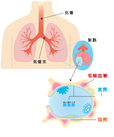
The lungs are made up of many small air-filled sacs called “alveoli”. The "interstitium" is the area between the alveoli and corresponds to the outer wall. In contrast, the portion within the alveoli is called the lung parenchyma.

What is the difference between normal pneumonia and interstitial pneumonia?

Diseases commonly called "pneumonia" are often caused by infectious diseases, in which bacteria proliferate in the alveoli (parenchyma) and cause inflammation. On the other hand, pneumonia in which the main pneumonia is in the wall (interstitium) of the alveoli is collectively called interstitial pneumonia. In other words, "interstitial pneumonia" is a name that distinguishes based on the location of pneumonia, and is simply called interstitial pneumonia.

However, the causes and course of the disease are different.
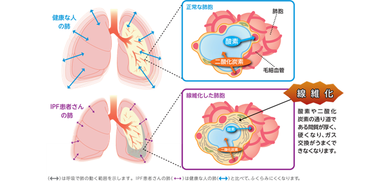
When interstitial pneumonia persists for a long period of time, ``fibrosis'' occurs in which the interstitium thickens and hardens due to the action of tissue repair. When fibrosis occurs in the interstitium, the lungs harden and cannot expand and expand sufficiently, leading to decreased vital capacity and shortness of breath. In addition, normal gas exchange is disturbed, and the oxygen concentration in the body tends to decrease even with a little movement.

What are the symptoms of interstitial pneumonia?

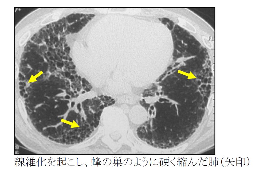
Cough and shortness of breath are the main symptoms. Those with rapid progress are often accompanied by fever. In long-lasting cases, fibrosis occurs, causing the lungs, which are originally soft like a sponge, to harden and shrink, leading to a decrease in vital capacity.

What is the course of interstitial pneumonia?

Interstitial pneumonia varies greatly in the speed and mode of progression for each type. In addition, even with the same diagnosis (eg, idiopathic pulmonary fibrosis), the course varies greatly from patient to patient. Some people have a gradual decline in respiratory function, while others have a rapid progression. Some people repeatedly experience a condition called acute exacerbation, in which the respiratory condition suddenly worsens over a short period of a few days to a month.

What is acute exacerbation of interstitial pneumonia?

Interstitial pneumonia is a group of diseases in which inflammation and fibrosis occur in the interstitium of the lungs, and respiratory function gradually deteriorates. Occasionally, however, a sudden exacerbation beyond the range of chronic progression occurs, and this is called an acute exacerbation. Specifically, it is a condition in which new shadows appear rapidly in the lungs within a few days to a month, leading to respiratory failure (a state in which necessary oxygen cannot be taken in). Patients with idiopathic pulmonary fibrosis are said to have an acute exacerbation at a rate of 5-15% per year.

In many cases, the cause of acute exacerbation cannot be identified, but acute exacerbation may occur after infection, surgery under general anesthesia, administration of new drugs (anticancer drugs, etc.), etc. Supplements and Chinese herbal medicines may cause acute exacerbation, so it is recommended that patients with interstitial pneumonia consult their doctor before starting any new medicines or supplements.

Acute exacerbation of interstitial pneumonia rapidly progresses to respiratory failure (inability to take in necessary oxygen). It is a serious condition with no established cure and a high mortality rate. Treatment involves the use of drugs such as steroids and immunosuppressants. Respiratory support, such as a ventilator, may also be needed. In addition, even after recovering from an acute exacerbation, there may be aftereffects on respiratory function, such as the need for home oxygen therapy.

2) How are interstitial pneumonias classified?

Identified cause
  • hypersensitivity pneumonitis
    Interstitial pneumonia caused by inhalation of various organic substances. It is caused by mold on housing, humidifiers, air conditioners, feed, etc., and dust from birds such as duvets and down products.
  • pneumoconiosis
    Interstitial pneumonia caused by inhalation of various inorganic substances. Examples include silicosis caused by inhalation of silicic acid compounds contained in rocks and asbestosis caused by inhalation of asbestos.
  • Interstitial pneumonia associated with connective tissue disease
    Interstitial pneumonia may occur with rheumatoid arthritis, scleroderma, dermatomyositis, polymyositis, Sjögren's syndrome, vasculitis, etc.
  • Radiation pneumonitis after radiotherapy
  • Drug-induced pneumonia caused by taking drugs
  • Interstitial pneumonia as an infection caused by viruses, some fungi, etc.
Those whose cause cannot be identified = idiopathic interstitial pneumonia (idiopathic is a medical term meaning that the cause is unknown)
  • chronic fibrosis
    ✓Idiopathic pulmonary fibrosis (IPF): the most common type
    ✓Idiopathic nonspecific interstitial pneumonia (NSIP)
  • smoking related
    ✓Desquamative interstitial pneumonia (DIP)
    ✓ Respiratory bronchiolitis-related interstitial lung disease (RB-ILD)
  • Acute/Subacute
    ✓Idiopathic organizing pneumonia (COP)
    ✓Acute interstitial pneumonia (AIP)
  • rare
    ✓Idiopathic pleural parenchymal fibroelastosis (PPFE)
    ✓Idiopathic lymphocytic interstitial pneumonia (LIP)

3) How is interstitial pneumonia diagnosed?

Opportunity to consult a medical institution

Interstitial pneumonia is often found in patients who visit a nearby hospital for dry cough or shortness of breath, and chest radiographs show abnormalities. A completely asymptomatic person may be diagnosed with an abnormality during a regular check-up and may be referred to a medical institution. If interstitial pneumonia is suspected, it is important to visit a medical institution capable of specialized diagnosis and treatment.

imaging test

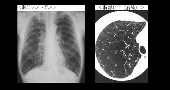
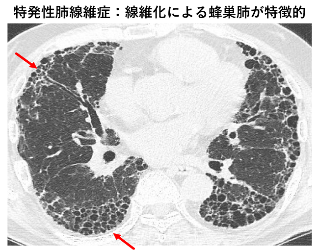
High-resolution CT (HRCT), which can take more precise lung images than normal CT, analyzes the characteristics of interstitial pneumonia in detail. This allows us to obtain information about the causes, types, and treatment of interstitial pneumonia. CT examination is useful not only for diagnosing interstitial pneumonia, but also for judging disease progression, therapeutic effect, and detecting complicating lung cancer.

Blood test

Markers such as KL-6 and SP-D, which are elevated in interstitial pneumonia, are measured and used for diagnosis and determination of therapeutic effects. If autoantibodies are elevated, it is likely that interstitial pneumonia is caused by an autoimmune disease. However, autoantibodies may mistakenly attack a part of the body as a foreign substance, causing interstitial pneumonia.)

respiratory function test

Interstitial pneumonia reduces lung capacity and gas exchange capacity. These things are measured using a machine called spirometry. It is also used to determine the effectiveness of treatment.

6 minute walk test

To evaluate the effect of interstitial pneumonia on cardiopulmonary function. Exercise endurance is assessed from the distance you can walk in 6 minutes. Since the examination is performed while wearing a pulse oximeter, it is possible to evaluate how much oxygen saturation in the blood changes due to exercise.

Tests to remove cells or tissues (bronchoscopy, lung biopsy)
bronchoalveolar lavage

A bronchoscope is used to wash the alveoli with saline and collect the washings. The type of disease is estimated from the components and cell types contained in the collected fluid.

forceps biopsy

A test in which small forceps are inserted through a bronchoscope to remove tissue from the periphery of the lung. Diagnosis in chronic interstitial pneumonia is limited due to the small tissue size available.

Frozen biopsy (cryo-biopsies)

This is a new method in which a special test instrument is inserted through the bronchoscope, the area around the tip is frozen, and tissue from the periphery of the lung is collected. It is becoming popular as a diagnostic procedure for interstitial pneumonia. The amount and quality of tissue that can be collected is superior to conventional forceps biopsy, and we are actively doing it in our department. You will be hospitalized for 3 days and 2 nights.

Thoracoscopic lung biopsy

A small hole is made in the body surface and lung tissue is collected from the outside of the lung using an endoscope called a thoracoscope. Since a large tissue is obtained, the amount of information is large, but it must be performed under general anesthesia like surgery.

Diagnosis of interstitial pneumonia is made by combining the above tests. The necessary tests differ depending on the type of disease, so please consult with your doctor before proceeding with the tests.

4) How is interstitial pneumonia treated?

treatment goal

The approach to treating interstitial pneumonia varies greatly depending on the type of interstitial pneumonia. For example, in the most common type of idiopathic pulmonary fibrosis (IPF), fibrosis-stiffened lungs often do not recover, making slowing future disease progression a realistic goal. On the other hand, in cases of collagen disease caused by autoimmunity and hypersensitivity pneumonitis caused by inhalation, some degree of recovery can be expected by removing the cause and suppressing inflammation. Before starting treatment, it is important to conduct sufficient examinations necessary for diagnosis and to set an appropriate treatment policy.

remove the cause

Interstitial pneumonia with a known cause is started by eliminating the cause as much as possible. If a drug is the cause, stop the drug. For interstitial pneumonia (hypersensitivity pneumonitis) caused by environmental allergens such as feathers and mold, remove or avoid the causative agent.

drug therapy

Drugs for treating interstitial pneumonia are divided into (1) drugs that suppress lung fibrosis (antifibrotic drugs) and (2) drugs that suppress inflammation (steroids and immunosuppressants).

  1. antifibrotic drug
    It is used for fibrosis-centered types of interstitial pneumonia. Two antifibrotic agents are currently approved for the treatment of idiopathic pulmonary fibrosis (IPF). Both Pirfenidone (Pirespa®︎, Pirfenidone®︎) and Nintedanib (Ofev®︎) have been shown to approximately halve the rate of lung capacity decline. It is also expected to suppress acute exacerbation of interstitial pneumonia. Furthermore, from 2020 onwards, nintedanib (Ofev®︎) will be available for interstitial pneumonia, which progresses fibrosis even with standard treatment, regardless of the type, increasing treatment options. spread.
  2. steroids, immunosuppressants
    Both are anti-inflammatory drugs and are used for interstitial pneumonia with strong inflammation. If long-term use is necessary, steroids and immunosuppressants may be used in combination. A typical target disease is interstitial pneumonia associated with an autoimmune disease (collagen disease). All of these drugs weaken your immune system, so you need to be careful about bacterial and viral infections while taking them. For acute exacerbation of interstitial pneumonia, large doses of steroids are administered to strongly suppress inflammation occurring in the lungs.
Other treatments

In order to minimize the impact of coughing and shortness of breath on daily life, it is important not only to treat interstitial pneumonia itself, but also to take care of exercise and nutrition to improve your overall condition.

  1. exercise therapy
    Shortness of breath is caused by the addition of three forces: lung power, heart power, and muscle power. When the power of the lungs is declining, it is important to continue appropriate physical activity in order to maintain and improve physical function. Walking is the main exercise that can be done at home. Learn how to move without straining your breathing.
  2. nutritional therapy
    When you are short of breath, you need more energy than usual to breathe. It is necessary to be careful of the vicious cycle of losing appetite and losing weight and becoming even more short of breath. Be conscious of "high calorie" and "high protein" and give due consideration to your diet.
  3. oxygen therapy
    It is performed when the lungs cannot take in enough oxygen due to deterioration of respiratory function. Install an oxygen concentrator at home to concentrate the oxygen in the air and inhale it through your nose. It is also possible to carry a portable cylinder and go out. There are several types, such as highly portable liquid oxygen and portable oxygen concentrators, so choose the one that best suits your medical condition and lifestyle.
  4. lung transplant
    If the disease progresses despite drug therapy, transplantation may be indicated. Lung transplantation includes brain-dead lung transplantation from brain-dead donors and living-donor lung transplantation from healthy donors. There are some conditions for lung transplantation, so please ask your doctor for details.
  5. clinical trial
    Clinical trials are conducted with the aim of confirming the efficacy and safety of a “candidate therapeutic drug” in healthy people and patients. Once a drug is approved by the government based on the results of clinical trials, it can be used by a large number of patients for the first time. Many drugs are still being developed for interstitial pneumonia, and our hospital is actively conducting clinical trials to improve the treatment results for interstitial pneumonia. Please ask your doctor or study coordinator for more information.

5) Start a What-if discussion (advance care planning)

In the unlikely event that the disease progresses, it is called "advance care planning" to think about the response and treatment in advance. As interstitial pneumonia progresses, shortness of breath may interfere with daily life. If you have any thoughts, please tell the medical staff and your family. We also want to support the feelings of our patients.

6) What medical systems are available for the treatment of interstitial pneumonia?

Intractable disease medical expenses subsidy system

This is a system that subsidizes medical expenses related to intractable diseases and reduces the out-of-pocket burden if the symptoms are more than a certain level or if you are paying high medical expenses for a long time among the "designated intractable diseases" specified by the government. Not all interstitial pneumonia is eligible, idiopathic pulmonary fibrosis (IPF) or idiopathic interstitial pneumonia other than IPF diagnosed by lung biopsy is designated as an intractable disease (specific disease). I'm here.

High-cost medical care benefit system

For those who are not covered by the Intractable Disease Medical Expenses Subsidy Program, there is a system in which the copayment of medical expenses is reduced by the health insurance (medical insurance) that you are enrolled in. Of the medical expenses incurred in the same month (from the 1st to the end of the month), the amount exceeding the copayment limit will be exempted or refunded. The maximum copayment amount depends on your health insurance and household income.

Long-term care insurance system

This system is available to those who wish to receive nursing care and support in their daily lives, and who are recognized as needing nursing care and support by the municipality. Nursing care services can be used by paying 10 to 30% of the cost of the service. Eligible people are those aged 65 or over who are recognized as requiring nursing care or support, and aged 40 to 64 who require nursing care or support due to 16 types of specified diseases (presenile dementia, cerebrovascular disorder, cancer, etc.). A person identified as needing assistance."

Handicapped person's notebook

Persons with physical conditions specified in the Act on the Welfare of Persons with Physical Disabilities can apply for this certificate. Disorders vary and may be subject to respiratory dysfunction when introducing home oxygen. Please check with your doctor to see if you are eligible. Depending on your grade, you may be able to receive a medical expense subsidy, and the details differ depending on the municipality where you live.

Daily life equipment supply business (subsidy for purchase of pulse oximeter)

This is a project that provides expenses for the purchase of equipment for the convenience of persons with disabilities in their daily lives. The content varies depending on the municipality where you live. In Kobe City, people with respiratory dysfunction level 3 or higher can receive payment for the purchase of a pulse oximeter. Please note that you must apply before purchasing.

7) Interstitial Pneumonia Support Team

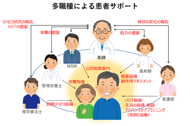
At our hospital, doctor, nurses, pharmacists, physiotherapists, nutritionists, social workers, and other multidisciplinary professionals collaborate with each other in their specialized approaches to treat these diseases, which are difficult to diagnose and treat. We are working to introduce treatment, maintain daily life, and improve quality of life. Specifically, doctor diagnose illnesses and explain treatment methods, nurses provide daily life guidance and support for home oxygen therapy, pharmacists provide detailed medication guidance, side effect management, physical therapists provide rehabilitation, nutritionists provide nutritional guidance, and social workers. effective use of social resources by In order to deepen these understandings, we have handed out the "Interstitial Pneumonia Handbook". We will also give you a booklet called "Palliative Care for Respiratory Failure" for patients whose disease has progressed and requires palliative care.

For reference, please download from the link below. (The copyright is reserved, so please refrain from reprinting to others without permission.)

Click here to download "Interstitial Pneumonia Handbook" 「間質性肺炎とどう付き合うか」のダウンロードはこちら

"I have trouble breathing" or "I can't breathe" are not the same as respiratory failure. The kanji character for "breath" is written as "one's own heart," but heart disease and mental problems can also make it difficult to breathe. Conversely, respiratory failure may occur even if breathing is not difficult. All animals, including humans, cannot live without oxygen. , defined as ``when the arterial blood gas partial pressure is less than 60 Torr during air inhalation''. Arterial blood gas partial pressure cannot be measured without drawing blood from the artery, but if you use a device called a pulse oximeter, you can measure the oxygenation level of arterial blood (arterial blood oxygen saturation SpO2) in real time simply by shining a light on your finger. can be measured and the arterial blood gas partial pressure can be easily estimated. An SpO2 of 90% or less corresponds to an arterial blood gas partial pressure of 60 Torr or less, so it can be said that there is respiratory failure. Pulse oximeters are also capable of continuous recording, and compact, lightweight ones are widespread, and are used in various places such as hospitals for respiratory management.

How does respiratory failure occur?

Breathing is the work of taking oxygen from the air into the body through the airways and lungs and expelling carbon dioxide gas from the body. Respiratory failure occurs when it becomes difficult to dissolve inside. However, even if there are no abnormalities in the respiratory tract or lungs, a neurological disease may weaken the respiratory muscles, a chest deformity may prevent the lungs from inflating sufficiently, or drugs may weaken the function of the brain's respiratory center. Respiratory failure may occur in some cases.

What types of respiratory failure are there?
Respiratory failure type I and respiratory failure type II

Respiratory failure is divided into two types: the type in which unnecessary carbon dioxide gas does not accumulate in the body (type I respiratory failure) and the type in which it accumulates (type II respiratory failure). Type I respiratory failure, also called pulmonary failure, includes asthma attacks, pulmonary fibrosis, and pulmonary embolism. Type II respiratory failure, also called ventilatory failure type, includes advanced pulmonary emphysema, pulmonary tuberculosis sequelae, thoracic deformity, and drug-induced respiratory depression.

Acute and Chronic Respiratory Failure

If respiratory failure persists for more than one month, it is classified as chronic respiratory failure, and if not, it is classified as acute respiratory failure. Patients with chronic respiratory failure require long-term treatment for respiratory failure, often exacerbated by a variety of causes. Emphysema, tuberculosis sequelae, pulmonary fibrosis, bronchiectasis, and thoracic deformities can cause chronic respiratory failure. On the other hand, acute respiratory failure rarely becomes chronic, and once the acute phase is overcome, treatment for respiratory failure is usually not required. Pneumonia, asthma attacks, drug intoxication, and pulmonary embolism can cause acute respiratory failure.

How is respiratory failure evaluated?

Although arterial blood gas partial pressure and arterial blood oxygen saturation (SpO2) can be measured with a pulse oximeter, the degree of respiratory failure at that time can be determined. Hugh-Jones's classification is a five-step method for indicating whether a country is

Hugh-Jones Classification
First degree normal
Second degree They can walk as well as healthy people of the same age, but cannot climb stairs or slopes as well as healthy people.
III degree They cannot walk on level ground like normal people, but they can walk more than 1.6 km (1 mile) at their own pace.
IV degree I can't walk more than 50m without resting.
V degree Shortness of breath even when talking or putting on or taking off clothes. I can't go out because I'm short of breath.

Pulmonary function tests and the 6-minute walk test (how far you can walk on flat ground in 6 minutes) may also be used to assess the degree of respiratory failure.

What is the treatment for respiratory failure?

Since there is a lack of oxygen in the body, treatment to replenish the body with oxygen is the first priority in any type of respiratory failure.

Airway management

If phlegm, blood, foreign objects, etc. are suffocating, first suction is used to remove them from the throat in order to secure a route to supplement oxygen. inserts a tube called a tracheal tube into the trachea through the nose or mouth.

oxygen therapy

Oxygen is usually inhaled through the nose or mouth through a cannula or mask, but the required flow rate of oxygen varies depending on the underlying disease and the degree of respiratory failure. In acute respiratory failure, a high flow rate (3-4 L/min) is sufficient from the beginning, but in chronic respiratory failure, in the case of type II respiratory failure in which carbon dioxide builds up in the body, high flow rate weakens ventilation further. I usually start with a low flow rate (3 L/min or less). Patients who cannot breathe easily on their own or who have severe respiratory failure are given artificial respiration, which pushes oxygen out through the mouth, nose, or tracheal tube.

artificial respiration

Artificial respiration can be performed by blowing air into the patient's mouth and nose as an emergency treatment, but in hospitals, a tracheal tube is usually connected to a respirator and oxygen is pushed into the trachea by a machine. However, since the insertion of a tracheal tube is extremely painful, recently, a method of artificial respiration using a nasal or nose-mouth mask instead of a tube (noninvasive ventilation therapy) has become widely practiced. I was.

drug therapy

Oxygen therapy and artificial respiration only make up for the lack of oxygen, so for patients with respiratory failure, oxygen therapy should be combined with antibiotics for pneumonia, steroid hormones and bronchodilators for asthma, etc. I also need treatment for my illness.

What is home oxygen therapy?

It is one of the most popular home therapies in Japan, and more than 100,000 chronic respiratory failure patients are currently receiving this treatment. It was in 1985 that I received social insurance coverage, and before that there were many patients who had to be hospitalized for a long time because they needed oxygen inhalation. Currently, the indication criteria for this treatment are those who have a resting arterial blood gas partial pressure of 55 Torr or less (88% or less by pulse oximeter SpO2), or a person whose arterial blood gas partial pressure is 60 Torr or less, but which drops significantly during exercise or sleep.
It is also indicated for patients with pulmonary hypertension and cyanotic heart disease. Many patients with chronic respiratory failure such as pulmonary emphysema, pulmonary tuberculosis sequelae, pulmonary pulmonary disease, and bronchiectasis receive this treatment, but recently, the number of lung cancer patients is also increasing. Stationary devices that concentrate oxygen in the air are often used to supply oxygen, but in this case, portable oxygen cylinders are usually used. There are also cases where liquid oxygen packed in a special container is used, but it is not as popular as a concentrator due to problems such as refilling and the location of the equipment. When you start home oxygen therapy, you tend to have a narrow range of activities, but you can actually travel, and the oxygen supply company will deliver oxygen concentrators and oxygen cylinders to your accommodation. In addition, there are groups of patients receiving home oxygen therapy in various places. However, once you start home oxygen therapy, it is important to have regular check-ups with your doctor to check your breathing.

What is home ventilation therapy?

At-home artificial respiration therapy is for patients with type II chronic respiratory failure in which carbon dioxide accumulates in the body. There is a way. The former, artificial ventilation through the tracheostomy, is mainly performed for patients whose respiratory muscles have become extremely weak due to neurological disorders. On the other hand, the latter, artificial respiration using a special mask, is often performed only at night in combination with home oxygen therapy for patients with chronic respiratory failure such as chest deformation, tuberculosis sequelae, and emphysema. In recent years, the number of patients has been increasing rapidly, partly because it is easy to use.

What about prevention of acute exacerbations?

Chronic respiratory failure often presents with an acute exacerbation requiring hospitalization, most commonly due to the common cold and subsequent respiratory tract infection. Although it is difficult to prevent this completely, it is important to maintain good hygiene such as gargling, brushing teeth, and washing your hands on a daily basis, as well as build up your resistance by eating a well-balanced diet, getting enough sleep, and exercising moderately. Influenza vaccines and pneumococcal vaccines are also preventive, so please consult your doctor. Other causes include chronic heart failure (cor pulmonale) from pulmonary hypertension due to chronic lack of oxygen, and even if this heart failure worsens, it will become acutely exacerbated. Excessive exercise, fatigue, and prolonged lack of oxygen, as well as excessive water intake, often cause swelling in the shins and puffy eyelids. For patients with cor pulmonale, it is also possible to prevent exacerbation by ``restricting water and salt intake, taking diuretics, increasing oxygen intake during physical activity, and avoiding excessive exercise''. increase.

Introduction…

Many of you have probably heard the term “sleep apnea”. In February 2003, it became famous for the incident of falling asleep while driving on the Sanyo Shinkansen, and has often been featured in newspapers and on television. In fact, it is closely related to not only drowsiness but also familiar diseases such as high blood pressure and heart disease. Sleep apnea is a very common disease, and it is estimated that about 20% of men and 10% of women over the age of 50 have sleep apnea to be treated. If you have any idea after reading the information below, why don't you visit a medical institution once?

What is sleep apnea

The majority of sleep apnea is "obstructive sleep apnea," where breathing stops (apnea) or nearly stops (hypopnea) during sleep. Subjective symptoms include snoring during sleep, inability to get a good night's sleep, waking up in the middle of sleep, headache upon awakening, drowsiness during the day, and loss of concentration. The person is often unaware, and family members often notice that breathing has stopped during sleep. Sleep apnea not only interferes with work and daily life and causes traffic accidents, but also causes various complications such as hypertension, arrhythmia, cerebral hemorrhage, and myocardial infarction. If it is severe, it is known that life expectancy will be shortened without treatment, and aggressive treatment is recommended.

Causes of sleep apnea

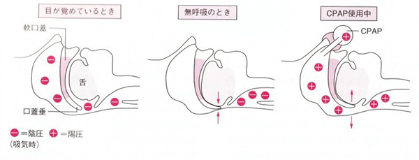
Obstructive sleep apnea is caused by partial or complete blockage of the airway, which is often the airway (see Figure 1). Obese people, people with short and thick necks, and people with small chins tend to have particularly narrow airways and are known to be prone to sleep apnea. Obese middle-aged and elderly men have the highest incidence of sleep apnea, and about half of those with metabolic syndrome have obstructive sleep apnea syndrome.

Diagnosis of sleep apnea

In principle, patients are hospitalized for one night, and their brain waves, oxygen concentration, chest movement, pulse, and airflow from the nose and mouth are measured during sleep (this test is called polysomnography), and the data is analyzed. Check the frequency of apnea and hypopnea during sleep. There is also a simple polysomnography that can be done at home.

Treatment of sleep apnea

In order to improve other lifestyle-related diseases such as blood pressure and diabetes, if you have obesity, you should work on weight loss first. However, in many cases, weight loss alone does not sufficiently improve apnea, and if there is more than a certain amount of apnea, treatment for the apnea itself is recommended. In addition, alcohol, sleeping pills, and anti-anxiety drugs relax the muscles of the pharynx, exacerbating airway obstruction.

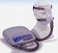
Nasal Continuous Positive Airway Pressure Device (CPAP) (see Figures 1 and 2)

It is a device that clears airway obstructions by sending air under a certain pressure through the nose, and is considered the first choice for the treatment of obstructive sleep apnea. If the number of apnea hypopneas per hour of sleep measured by polysomnography is 20 or more (40 or more for the simple type), insurance will be applied.

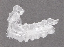
Oral appliance (mouthpiece) (see Figure 3)

A device that is fixed between the upper and lower teeth to prevent the lower jaw from sinking, which is one of the causes of upper airway obstruction. Have your doctor make one. It works primarily in patients with mild to moderate obstructive sleep apnea.

surgery

Surgery is a good indication if the primary cause of airway obstruction is an enlarged tonsil or adenoids in the absence of obesity, but primarily for sleep apnea in children.

At the end…

If you have any of the following symptoms, you may have sleep apnea, so we recommend that you see a doctor. In particular, people with lifestyle-related diseases such as obesity, high blood pressure, and diabetes, as well as those with heart and blood vessel diseases, need to be careful.

  • Snoring is said to be terrible. I hold my breath sometimes.
  • Feeling sleepy during the day, even though you should be getting enough sleep.
  • I often wake up during the night. I feel suffocated.

COPD is an acronym for Chronic, Obstructive, Pulmonary, and Disease. Inflammation caused by smoking causes chronic cough, phlegm, and shortness of breath due to alveolar wall destruction and bronchial stenosis. It is a disease that causes

Causes of COPD

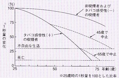
In the past, air pollution was also a cause of COPD, but in today's developed countries most of the causes are smoking. People who have never smoked rarely develop COPD, so it is true that smoking is the cause of COPD, but not all smokers are the cause of v, and about 15% of smokers develop COPD. However, the remaining 85% of smokers will not develop COPD. Therefore, it is thought that there are factors other than tobacco smoking that are responsible for tobacco susceptibility (susceptibility to emphysema). However, even today, the differences between those who develop emphysema and those who do not are unknown and cannot be predicted. The only way to avoid COPD is to stay away from cigarettes. One prediction states that if you quit smoking by age 45 you can avoid COPD, but if you quit smoking after age 65 you will no longer be able to avoid COPD. If a person with COPD does not stop smoking as soon as possible, COPD will progress. The most important thing after being diagnosed with COPD is to stop smoking immediately.

Symptoms of COPD

It begins with symptoms such as coughing and phlegm, and gradually develops shortness of breath when moving (especially when climbing stairs), and eventually dyspnea occurs even with mild daily activities. In severe cases, a large amount of energy is used for breathing, which can lead to weight loss, muscle atrophy, and decreased physical strength, leading to bedridden conditions.

Diagnosis of COPD
A pulmonary function test (a test to breathe in and out like vital capacity) measures vital capacity in 1 second (the amount exhaled in the first 1 second), and the airway narrows and it takes time to exhale. can be largely diagnosed by detecting , and is an indicator of severity. In mild cases, COPD may appear normal on x-rays, but in severe cases, the lungs may appear excessively inflated. In addition, high-resolution chest CT can detect lung destruction and detect early-stage emphysema.
Treatment of COPD
no smoking

There is no way to repair the already damaged parts of the lungs and airways. The first priority is to stop smoking immediately so that it does not progress any further. This is because even if you take medication while smoking, you cannot avoid getting worse. At the Smoking Cessation Outpatient, you can quit smoking under the guidance of a doctor using smoking cessation aids (alleviating nicotine withdrawal symptoms).

drug therapy

The most effective treatment is smoking cessation and inhalation of bronchodilators (anticholinergics, long-acting β2 agonists). In severe cases and repeated exacerbations, inhaled steroids are used in combination. Currently, there are multiple long-acting anticholinergics, β2 agonists, inhaled steroids, and combinations of these on the market, and the appropriate inhaler is selected for each individual patient. These inhalations do not have an immediate effect, and it is important to inhale regularly every day, not when you are sick. It increases the amount of exercise you can endure (that is, you become able to do things you couldn't do because you're short of breath) and improves your quality of life, but it's not thought to prolong your prognosis (how many years you can expect to live). increase.
In addition, expectorants and steroids are used in severe cases.

oxygen therapy

If it progresses, home oxygen therapy (a treatment in which an oxygen machine is installed at home and oxygen is inhaled at home or during exercise) is necessary to compensate for the lack of oxygen. Home oxygen therapy has been shown to prolong prognosis. Carbon dioxide builds up in the blood and may require a mask-type ventilator.

rehabilitation

In addition to respiratory failure, COPD is also a systemic inflammatory disease that results in loss of muscle mass and nutritional deficiencies. Exercise therapy, such as lower extremity muscle strengthening and pulmonary rehabilitation, has an additional effect on drug therapy. In addition, weight loss is said to increase the risk of progression to respiratory failure and death, so nutritional therapy to prevent weight loss is important.

acute exacerbation

It is known that acute exacerbation of COPD can lead to aggravation of pneumonia and bronchitis, and the frequency is higher in severely ill people. This may be the case, for example, if shortness of breath develops rapidly. Most cases require hospitalization and have a high mortality rate. Expectorants, macrolide antibiotics, vaccines (influenza, pneumococcus), etc. are said to be effective in preventing exacerbation.

Prognosis of COPD

The 5-year survival rate (indicator of how many people are alive after 5 years) after the appearance of shortness of breath is about 70%, and the 10-year survival rate is about 40%. The prognosis is particularly poor in the elderly and those who continue to smoke. Patients with COPD are more likely to develop lung cancer later in life.

At the end

If you are 45 years of age or older, have smoked for 10 years or more, and have cough, sputum, or shortness of breath on exertion, we recommend that you see a doctor once.

Asthma (asthma) is a chronic disease of the "bronchi", which is the passage of air. The airways of people with asthma are chronically inflamed, making them more sensitive to stimuli (called airway hyperreactivity). An "asthmatic attack" occurs when the bronchi become narrower in response to stimulation, and as you breathe through the narrowed bronchi, you will hear a wheezy whistling sound and feel suffocated. Asthma is characterized by bronchial narrowing that resolves with treatment or spontaneously (reversible airflow limitation). Asthma is a disease with strong symptoms, but because it is easy to get used to the symptoms, it is difficult to correctly diagnose the condition, and as a result, it is often the case that appropriate treatment is not provided. To properly treat asthma, it is first necessary to understand what asthma is.

What is airway inflammation?

The airways of people with asthma are chronically inflamed. It was known that cells called eosinophils were involved in inflammation, but recently it has been found that other inflammatory cells such as lymphocytes and neutrophils, as well as various substances, are also involved. rice field. Inflammation persists even when there are no symptoms, so treatment must be continued outside attacks. (described later)
To see the degree of inflammation, the number of inflammatory cells in sputum and the concentration of a substance called nitric oxide (NO) in exhaled breath are measured.

What is airway hyperreactivity?

An asthma patient's bronchi are inflamed and swollen, just like a burn. Just as a burn hurts to the slightest touch, an asthmatic's bronchial tubes become hypersensitive to stimulation. (This is called airway hyperreactivity). The airway narrows in response to things that may not be irritating to the average person, such as cigarette smoke, pollen, or cold air.
The degree of airway hyperresponsiveness can be assessed using substances that constrict bronchos (such as methacholine) (Methacholine Airway Hyperresponsiveness Test).

What is reversible airflow limitation?

Narrowing of the bronchi restricts airflow. This is called airflow restriction. Reversal of airflow limitation in patients with asthma has been characterized by treatment. This is called reversible airflow limitation. In recent years, it has been found that airflow limitation in asthma patients gradually becomes fixed. In other words, the bronchi gradually harden and narrow. (This is called remodeling.) Simultaneously with the prevention of attacks, prevention of progressive airflow limitation is considered an important future point in asthma treatment.

How many people have asthma?

It is the most common respiratory disease, with an estimated prevalence of 3-5 per 100 people. The prevalence of asthma is increasing year by year. In addition, many people develop asthma for the first time in middle-aged and elderly people, and in recent years, asthma in the elderly has been attracting attention.

What are the symptoms of asthma?

Symptoms of asthma include:

  1. Shortness of breath, shortness of breath (dyspnea)
  2. Wheezing, wheezing, and wheezing in the throat and chest
  3. Cough that lasts for days (can be productive or dry)

Not all people with asthma have wheezing or difficulty breathing. In some people, coughing is the only symptom of asthma (coughing asthma). Coughing in asthma often occurs at night or early in the morning, and during exercise, and wheezing and chest symptoms tend to intensify at night and early in the morning. Even very mild asthma symptoms can get worse if left untreated. Treating symptoms while they are mild can prevent symptoms from getting worse over time.

why do you get asthma

The underlying causes of asthma are still poorly understood. Asthma patients often have family members with asthma, and it is known that some kind of genetic predisposition is involved. Advances in genetic analysis in recent years have led to the elucidation of some of the genes related to asthma, but it will take more time to clarify the whole picture.

Are there types of asthma?

There are two types of asthma: those with strong allergies to house dust, mites, pollen, etc. (called atopic asthma) and those with poor atopic constitution (called non-atopic). In recent years, the diversity of asthma has been emphasized and various other classifications have been attempted.

1. Atopic asthma

It is a type of asthma that causes allergies to various organic substances in the indoor environment and outdoors. Atopic dermatitis and allergic rhinitis are likely to be complicated, and avoidance or elimination of allergens may be effective.

2. non-atopic

A type in which the allergen that triggers an asthma attack cannot be identified.

What are the signs of a dangerous seizure?

It is necessary to see a doctor before any of the following symptoms occur. If you do, you need to go to the hospital immediately (in some cases, by ambulance).

  1. Need to catch your breath during a conversation, can't speak loudly
  2. The inhalers (bronchodilators) that you usually use do not work well or do not work
  3. Mild seizures that occur daily
  4. If you have recently had a severe seizure (hospitalized or rushed to the emergency department)
  5. When you can't go to the toilet or when you become incontinent
  6. When you continue to be in pain and unable to lie down
Is asthma curable?

Unfortunately, there is currently no cure for asthma. However, if adequate treatment is given early after onset, most asthma patients can be completely controlled. Although some patients may be able to stop treatment altogether, there is always the possibility that symptoms will recur. On the other hand, it is believed that if the symptoms of asthma are left untreated, inflammation will destroy the tissues of the bronchi and lungs, causing a gradual loss of function that will never recover. When this happens, treatment cannot be stopped, and even with treatment, the symptoms do not improve sufficiently. Appropriate early treatment is important.

Please tell me about medicine for asthma

Medications for asthma are divided into long-term control drugs that are used daily and anti-attack drugs that are used as needed during an attack.

long-term control medication

Inhaled steroids are the mainstay of long-term control drugs. Steroids are drugs that can most effectively suppress airway inflammation, which is the essence of asthma, and can be used safely with almost no systemic side effects when used by inhalation. Daily inhalation suppresses airway inflammation, improves airway hypersensitivity (mentioned above), and prevents attacks.

When inhaled steroids alone cannot control

吸入ステロイドと長時間作動型のβ2刺激薬の合剤を用います。その他、ロイコトリエン受容体拮抗薬、テオフィリン製剤、長時間作動型の抗コリン薬といった薬を併用します。吸入ステロイドや併用薬を十分に使用してもコントロールできない重症喘息に対しては、近年抗IgE抗体や抗IL-5抗体といった生物学的製剤が使えるようになりました。

Seizure medication

The main treatment for seizures is the inhalation of short-acting bronchodilators (short-acting β2 agonists). Inhaled steroids are inhaled daily, and short-acting β2 agonists are used as needed when symptoms appear. For more severe attacks, steroids may be taken orally or given as an IV. However, the original goal of asthma treatment is to control asthma so that it does not require anti-seizure drugs in usual treatment.

Are there any factors that cause the control to fail?

If you know the allergen that triggers an asthma attack, try to avoid or eliminate the allergen. Smoking is strictly prohibited. Please observe the no-smoking policy. Allergic rhinitis, sinusitis, gastric acid reflux (reflux esophagitis), obesity, sleep apnea syndrome, etc. are known to cause poor control of asthma. Psychological factors such as excessive stress are also known to increase the risk of exacerbation of asthma. In this way, comprehensive management, such as evaluation and treatment of complications, allergen avoidance, and smoking cessation guidance, is considered important in asthma management.

Are asthma medications safe?

Inhaled corticosteroids and β2-stimulatory inhalants are the mainstays of treatment, but both have very few systemic side effects and pose no particular safety concerns. Safe even during pregnancy. The most important thing during pregnancy is not to have an asthma attack. It is said that when a seizure occurs, oxygen is not sufficiently distributed, which is the most damaging to the fetus.

Can I live a healthy life with proper treatment?

With proper treatment, many patients are able to:

  1. You can participate in various exercises and sports and live an active life without asthma symptoms.
  2. I can sleep without worrying about asthma symptoms.
  3. It can also prevent asthma attacks during the season of asthma exacerbation or when a trigger is encountered.
  4. Your lungs will function well and you will be able to maintain good lung function.

In other words, you can live a normal life like a healthy person.

Pneumonia is one word, but there are various types of pneumonia.

Bacterial pneumonia, viral pneumonia, drug-induced pneumonia, autoimmune disease pneumonia, and so on.
Among them, pneumonia generally refers to bacterial pneumonia, that is, pneumonia caused by germs. (For other types of pneumonia, please refer to the section on interstitial pneumonia.)
A long time ago, many people lost their lives due to pneumonia when there were no antibiotics. With the development of drugs, it is often cured by outpatient treatment. However, it can sometimes be severe and is still an important cause of death, especially in the elderly.

Which microbe is the causative agent?
Bacteria that cause pneumonia in outpatients

Bacteria with names such as Diplococcus pneumoniae, Haemophilus influenzae (not the influenza virus that causes the flu in winter), Staphylococcus aureus, Moraxella catarrhalis, and Mycoplasma are often the culprits.
In addition, it is reported that there are many viruses such as influenza virus (this is the person responsible for causing the famous influenza). Viruses and bacteria are the same microorganisms, but they are different in size and propagation method, so please think of them as completely different things.
In clinical practice, the causative pathogen is often unknown.

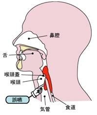
In addition, in patients who are elderly, have a history of stroke, or have underlying diseases such as those who have undergone stomach or esophageal surgery, chronically present in the mouth, throat, stomach, esophagus, and other gastrointestinal tracts. It is common for germs to enter the airways and cause recurrent episodes of pneumonia.
What are the symptoms?

It often shows so-called cold symptoms such as fever, cough, and phlegm. Other symptoms such as chest pain, general malaise, and difficulty breathing may also occur. These symptoms are not specific to pneumonia, and symptoms alone cannot be used to diagnose pneumonia.

what about the test?

A chest x-ray (sometimes a chest CT scan) and blood tests are needed to make a diagnosis of pneumonia. Microscopic examination and culture of sputum are also performed to find out what pathogen is the cause.

What about treatment?

Administration of antibiotics against the causative bacteria is the basis of treatment. Of course, rest, hydration and nutrition are important.
If it is mild, outpatient treatment is possible, but if outpatient treatment is ineffective, if systemic symptoms such as dehydration are severe, or if breathing is severe, you may need to be hospitalized and receive IV treatment or oxygen inhalation. . This is often the case with the elderly or those with underlying medical conditions.

Can we prevent it?

There is no sure-fire way to prevent this.
However, doing what everyone knows, such as avoiding crowds and practicing good hand hygiene, is especially important for patients with weakened immune systems.
Patients with chronic aspiration should not lie down for 1-2 hours after eating and should not eat or drink for 1-2 hours before going to bed. Keep the inside of the mouth as clean as possible by using mouthwash to reduce the growth of bacteria in the mouth, and if possible, ask the patient to sleep with the upper body slightly upright. This will reduce the risk of aspiration.
In addition, as mentioned above, the elderly and those with some underlying diseases are more likely to become seriously ill, so please consult your doctor about vaccination against influenza and pneumococcus, which is the most common cause of pneumonia.

Medical record

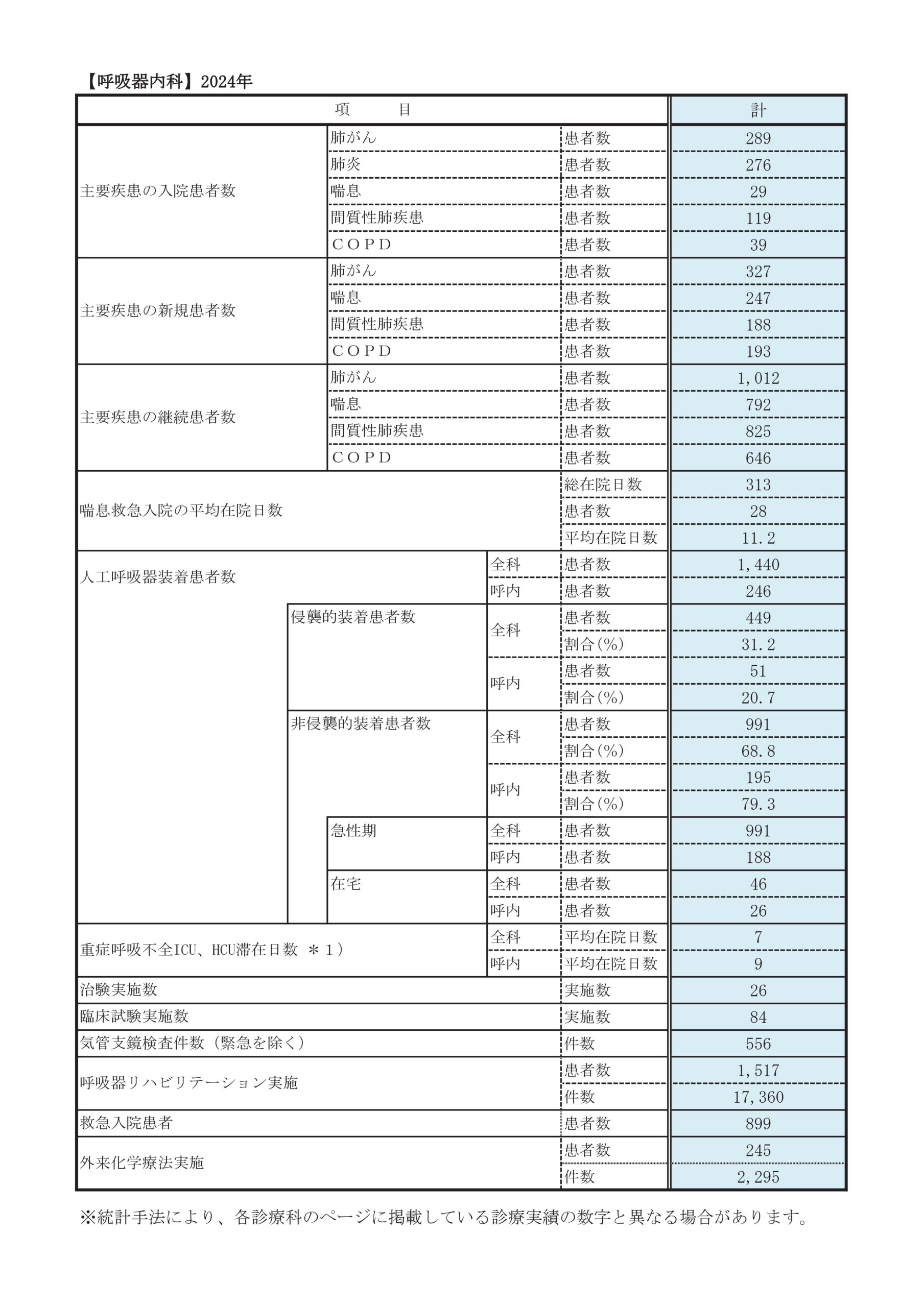
Departmental statistics

Clinical Metrics Page

clinical research

Research Project Name Principal Investigator Research implementation period attached file
Rehabilitation for patients with chronic obstructive pulmonary disease after hospitalization for exacerbation
Shirakawa Chikusa October 2024~
March 2028
PDF
A multi-regional cohort study aimed at generating evidence to improve residents' health: the LIFE Study Shirakawa Chikusa October 2024~
March 2028
PDF
A retrospective observational study on the relationship between pre-hospital frailty and prognosis in patients hospitalized with acute exacerbation of fibrotic interstitial pneumonia Ryo Tachikawa October 2024~
December 2026
PDF
A multicenter study on the use and efficacy of home high-flow nasal cannula oxygen therapy (HFNC) Keisuke Tomii April 2024~
March 2025
PDF
Investigation of infectious viruses and clinical features by multiple PCR testing in adult patients visiting the emergency room with respiratory exacerbations after the COVID-19 epidemic Takeshi Sasada February 2024~
March 2026
PDF
A retrospective study on the efficacy and safety of platinum-based etoposide plus PD-L1 inhibitors in patients with recurrent limited-stage small cell lung cancer after chemoradiotherapy Sato Yuuki From December 2023
March 2025
PDF
Construction of a database of lower respiratory tract infections in acute care general hospitals in Japan and evaluation of the effectiveness of empirical use of antibiotics with anti-Pseudomonas aeruginosa activity for recurrent lower respiratory tract infections: a multicenter retrospective cohort study (multi-institutional collaborative study) Keisuke Tomii November 2023~
December 2027
PDF
Multicenter collaborative study on establishing a diagnostic method for predicting the efficacy of cancer immunotherapy based on immune response analysis in solid cancers Keisuke Tomii November 2023~
January 2024
PDF
Cost-effectiveness study of anticancer drugs using real-world data (RWD) Comparative study of immunotherapy (Atezolizumab vs. Durvalumab) in extensive-stage small cell lung cancer Sato Yuuki October 2023~
December 2024
PDF
Nationwide epidemiological survey of hypersensitivity pneumonitis Keisuke Tomii From September 2023
March 2027
PDF
A multi-center, retrospective observational study evaluating the efficacy and safety of Ramucirumab + Erlotinib for advanced, recurrent non-squamous non-small cell lung cancer positive for EGFR gene L858R mutation (REAL-SPEED) Sato Yuuki From August 2023
December 2026
PDF
A case of ALK-positive lung cancer in which brain metastases after Alectinib and Brigatinib treatment were successfully treated with Lorlatinib Sato Yuuki From August 2023
July 2024
PDF
A retrospective observational study on the relationship between early treatment response and prognosis in patients with acute exacerbation of idiopathic pulmonary fibrosis treated with steroid pulse therapy Kazuma Nagata From August 2023
March 2026
PDF
A retrospective observational study of small cell lung cancer with paraneoplastic neurological syndrome Sato Yuuki

From June 2023
May 2024

PDF
A case report and literature review of EGFR-positive lung adenocarcinoma in which ICI monotherapy was effective only in the transformed lesions to squamous cell carcinoma Sato Yuuki From June 2023
May 2024
PDF
Nationwide survey of bronchiectasis associated with rheumatoid arthritis Keisuke Tomii February 2023~
March 2025
PDF
A retrospective study of patients undergoing chemoradiotherapy for locally advanced non-small cell lung cancer (HOPE-005/CRIMSON) Sato Yuuki February 2023~
January 2028
PDF
Immune microenvironment and clinical course of histological transformation in patients with EGFR mutation-positive lung cancer (HISTORIC-TR) Sato Yuuki January 2023~
May 2025
PDF
Observational study of patients with non-small cell lung cancer treated with nivolumab monotherapy Sato Yuuki From November 2022
December 2024
PDF
The relationship between early rehabilitation and rehospitalization in patients aged 65 years or older with chronic obstructive pulmonary disease who require nursing care level 1 or higher Keisuke Tomii From July 2022
March 2027
PDF
Investigation into the significance of TP53 gene mutations and the mechanism of treatment resistance in advanced and recurrent non-squamous non-small cell lung cancer positive for EGFR gene L858R mutation (WJOG14420LTR) Sato Yuuki February 2022~
February 2028
PDF
Survey project on biomarker testing and targeted therapy for advanced and recurrent non-small cell lung cancer Sato Yuuki January 2022~
December 2025
PDF
A study to establish a diagnostic method for the extent of empyema, which is useful for selecting treatment methods for empyema Keisuke Tomii From September 2021
December 2024
PDF
A retrospective study of lung cancer patients with rare driver gene alterations Sato Yuuki From July 2021
March 2025
PDF
Analysis of COVID-19 pathology and elucidation of the mechanism of aggravation Keisuke Tomii From June 2021
January 2026
PDF
Observational study on the treatment status, efficacy and safety of nivolumab + ipilimumab + chemotherapy combination therapy for untreated advanced or recurrent non-small cell lung cancer in Japan (LIGHT-NING) Sato Yuuki From December 2021
December 2025
PDF
Research into identification of prognostic predictors for COVID-19 and their clinical application Keisuke Tomii September 2020~
March 2025
PDF
Elucidation of the mechanism of aggravation of COVID-19 infection Keisuke Tomii September 2020~
March 2025
PDF
A multicenter survey of drug-induced lung injury associated with osimertinib, a first-line treatment for EGFR mutation-positive non-small cell lung cancer Sato Yuuki September 2020~
March 2025
PDF
Clinical and imaging features of COVID-19 Sato Yuuki September 2020~
March 2025
PDF
Epidemiology and biomarkers of diffuse lung disease Keisuke Tomii August 2020~
September 2029
PDF
Biomarker study accompanying a multicenter prospective observational study examining the efficacy and safety of first-line carboplatin/etoposide/atezolizumab combination therapy in clinical practice for patients with extensive-stage small cell lung cancer [APOLLO-Bio] Keisuke Tomii March 2020~
September 2025
PDF
Survey on histological transformation in EGFR mutation-positive lung cancer patients [HISTORIC] Sato Yuuki May 2020~
May 2025
PDF
Registry study on COVID-19 Jun Nakagawa May 2020~
March 2026
PDF
A multicenter survey of initial treatment with osimertinib for EGFR mutation-positive non-small cell lung cancer Sato Yuuki April 2020~
March 2025
PDF
A multicenter study on the clinical features of coronavirus pneumonia (CoVP), influenza virus pneumonia (InVP), and non-viral community-acquired pneumonia (nVCAP) Keisuke Tomii April 2020~
April 2025
PDF
A prospective multicenter collaborative study using the SCRUM-Japan disease registry to generate clinical trial control group data for new drug approval reviews [SCRUM-Japan Registry] Keisuke Tomii February 2020~
March 2026
PDF
Survey of intractable asthma complicated by bronchiectasis Keisuke Tomii February 2020~
December 2024
PDF
A retrospective study of patients who underwent chemoradiotherapy for locally advanced non-small cell lung cancer Sato Yuuki November 2019~
March 2025
PDF
Observational study on the long-term safety and efficacy of durvalumab after concurrent chemoradiotherapy in patients with unresectable stage III non-small cell lung cancer [AYAME study] Sato Yuuki September 2019~
December 2024
PDF
A multicenter observational study on when to stop molecular targeted therapy in patients with advanced lung cancer Sato Yuuki August 2019~
March 2027
PDF
Epidemiology of pneumococcal pneumonia in adult community-acquired pneumonia (COP) Keisuke Tomii August 2018~
March 2025
PDF

news

Introduction of our department for doctor and those who aspire to become doctor

*This content is intended for doctor and is intended to deepen the understanding of this medical institution, and is not intended for publicity or advertising for the general public. Nice to meet you, my name is Keisuke Tomii Tomii Respiratory Medicine Kobe City Medical Center General Hospital.
In our department, under the mission of a regional medical support hospital, we are responding to all respiratory diseases 24 hours a day, 365 days a year. is our motto.
Among them, today, we will introduce the latest medical treatment for lung cancer and interstitial lung disease supported by a multidisciplinary medical team, which is a feature of our hospital.

Keisuke Tomii
Vice President
Respiratory Medicine Director

(1) About lung cancer
Characteristics of lung cancer treatment at our hospital

Lung cancer is the number one cancer-specific cancer death toll in Japan in 2019, and it is a disease with a poor prognosis.

Our hospital is a regional cancer treatment base hospital representing the Hanshin area.

  1. Delivering appropriate standard treatment based on wide-ranging cooperation between clinical departments that utilizes the strengths of a general hospital
  2. Bridging to new treatments through cancer gene panel testing and clinical trials/clinical trials

We are working together to provide lung cancer treatment with the aim of becoming a hospital where local patients and family doctors can receive consultations and referrals with peace of mind.

Optimal treatment selection according to the stage of progression and improvement of patient QOL through team medical care

For early-stage lung cancer, where the cancer focus remains in the thorax, we aim for radical cure by making full use of a combination of surgery, radiation therapy, and chemotherapy, considering all possibilities. At our hospital, we have departments of respiratory Respiratory Medicine, Respiratory Sugery, and Radiation Oncology that are involved in the treatment of respiratory cancer. I will try to provide it in a timely manner.

On the other hand, advanced lung cancer with distant metastasis is mainly treated with chemotherapy. In addition to conventional cytocidal anticancer drugs, recently, immune checkpoint inhibitors targeting PD-1/PD-L1 and CTLA-4, and specific inhibitors of cancer-causing genes A molecular-targeted drug is used. EGFR and ALK are well-known molecular-targeted drugs, but in addition to these, inhibitors against ROS1, MET, and BRAF V600E are now available under health insurance. By making full use of these treatments, more patients are now able to maintain long-term survival and QOL, which was unimaginable until a decade ago.

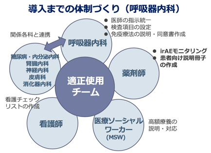
On the other hand, the side effects of anticancer drugs are diverse. In particular, the side effects of immune checkpoint inhibitors are called immune-related adverse events (irAE), which require skill in detection and post-onset management. may be required. At our hospital, we have created a proper use team that transcends the boundaries of related clinical departments and departments, and perform systemic management that makes the most of the strengths of a general hospital. In addition, our hospital has one of the leading emergency medical care centers in Japan, so you can feel free to consult us in the event of an emergency. In addition, the in-hospital palliative care team is also active, so it is possible to provide appropriate palliative care in parallel with treatment. If the situation leads to the maintenance of QOL, palliative radiation therapy and drainage for pleural effusion are also available.

Patient Benefits from Providing Advanced Cancer Treatment

As part of advanced cancer treatment, we are conducting a large number of clinical trials and clinical trials in all stages from early stage to advanced stage. For patients, there are significant benefits, such as access to new drugs and treatments and, in some cases, reduced medical costs. Recently, the term precision medicine has become popular, and drug therapy for lung cancer, in particular, is leading the way in cancer treatment. In fact, when detailed genetic testing is performed on non-small cell lung cancer patients, half of the patients are found to have some kind of genetic mutation, making it possible to use molecular-targeted drugs under medical insurance coverage.​ ​In addition, since our hospital is a cancer genome medical cooperation hospital, we can conduct cancer gene panel tests on patients who have completed standard treatment and examine whether there are therapeutic drugs that can be expected to be effective. In this case, tissue collection of cancer cells is important, and our hospital is very proactive in tissue biopsies such as ultrasound-guided bronchoscopy and CT-guided biopsy.

If you have a suspected patient with lung cancer, please introduce us first.

About half of advanced-stage lung cancers are asymptomatic, so it is okay to start immediately after an X-ray is suspected. Even elderly patients and patients with comorbidities may be actively indicated for treatment depending on the status of gene mutations and PD-L1, so please introduce us first. We also actively accept referrals for the purpose of cancer gene panel testing. Lung cancer became resistant to immunotherapy, molecular targeted drugs, and two types of cytotoxic anticancer drugs, while thymic tumors and malignant pleural mesothelioma became resistant to the first type of cytotoxic anticancer drug. The timing is considered to be a good timing for cancer gene panel testing. We would appreciate it if you could introduce us to the Cancer Genome Testing Outpatient Department (Tuesday afternoon) at our hospital if you are an applicable patient and wish to have a cancer gene panel test.

(2) Interstitial lung disease

Interstitial lung disease is a general term for diseases that cause inflammation and fibrosis in the lung interstitium. The rate of disease progression varies from acute (days) to chronic (years), with cough and shortness of breath being the predominant symptoms. There are various causes such as infection, smoking, allergies, autoimmunity, drugs, etc. First, the cause is identified and treated (removed).

Idiopathic pulmonary fibrosis, which is the most common chronic interstitial lung disease, has a poor prognosis of 3-5 years after diagnosis due to progressive fibrosis. I had no choice but to watch the progress while doing it. However, with the recent advent of antifibrotic drugs, it is expected that the progression of the disease will be suppressed and the prognosis will be improved.
Diagnosis of interstitial lung disease jointly with diagnostic Diagnostic Radiology and Clinical Pathology

Interstitial lung disease is an intractable disease that is particularly difficult to diagnose among various respiratory diseases. Our department has a wealth of experience in treating interstitial lung disease, and many patients visit us. Diagnosis requires comprehensive evaluation of medical history, physical findings, blood tests, HRCT findings, and bronchoscopic and thoracoscopic lung biopsy results. Therefore, we discuss with not only the Respiratory Medicine, but also the Department of Diagnostic Radiology and Diagnostic Clinical Pathology to determine the diagnosis and treatment policy.

In our department, we regularly hold study sessions with doctor from diagnostic Diagnostic Radiology and Clinical Pathology that specialize in interstitial lung disease nationwide, and discuss past cases to improve the accuracy of diagnosis. Recently, bronchoscopes have introduced frozen biopsies (cryo-biopsies), which can collect larger tissue than conventional forceps biopsies, making it possible to obtain pathological information more easily.

New drug therapy (antifibrotic drug) for interstitial lung disease

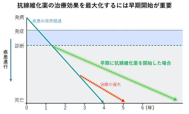
For idiopathic pulmonary fibrosis, two types of antifibrotic drugs, pirfenidone (Pirespa®) and nintedanib (Ofev®), are covered by insurance and are the main drug therapy. These agents have been shown to slow the progression of fibrosis, reduce the frequency of acute exacerbations, and improve prognosis. Recently, nintedanib has become available for interstitial lung disease other than idiopathic pulmonary fibrosis when progressive fibrosis is present. However, if the disease progresses and the lung capacity is severely reduced, it is difficult to improve symptoms and lung capacity even with these drugs. Therefore, it is considered important to diagnose as early as possible and start these drugs at the appropriate time to prevent progression. In addition, the clinical trial of the third antifibrotic drug is ongoing at our hospital.

Patient support by a multidisciplinary team

Progressive interstitial lung disease can cause shortness of breath, decreased activity, and weight loss as lung function deteriorates, even with the best treatment. In advanced stages, rehabilitation and palliative treatment to maintain function may be more important than drug therapy. In our department, we have established a system that allows multidisciplinary intervention even in the outpatient department, and specialists work together to support patients, such as comprehensive respiratory rehabilitation, lifestyle guidance, advanced care planning, and public system applications.

Early diagnosis and early treatment are the key to interstitial lung disease!

It is important to diagnose interstitial lung disease as early as possible and start treatment at an appropriate time. In the midst of the corona crisis, our department is providing medical care while devising measures to prevent infection, such as rehabilitation classes using the web. If a patient complains of coughing or shortness of breath, or finds even a slight opacity on a chest photo or CT, we would appreciate it if you could refer us to our hospital through the Community Liaison Office without hesitation.

About the medical treatment system under the new coronavirus epidemic at our hospital

Since March 2020, a new type of coronavirus infection has been prevalent in Hyogo Prefecture, and as a designated hospital for infectious diseases, our hospital is working day and night to respond mainly to severe cases. We are taking the maximum possible measures, such as setting up a dedicated ward and pre-hospital PCR testing, to prevent in-hospital infections and maintain regular medical care.

The battle against the new coronavirus is still ongoing, but people are reluctant to undergo medical examinations due to the epidemic, and there is a strong concern that the number of cancer patients will increase in the future.

In addition, in the midst of the corona crisis, it is difficult to provide rehabilitation guidance face-to-face, so our department is conducting respiratory rehabilitation classes using the web. You can receive lectures and practical guidance from our doctor and physical therapists using your home computer or smartphone. At our clinic, we will continue to explore all possible treatments and respond to the needs of our patients, such as relieving symptoms and maintaining quality of life, and will continue to develop the best treatment that will never be abandoned, so please feel free to contact us. Please give me.

Study session for regional medical cooperation

Kobe City Medical Center Diffuse Lung Disease Study Group

It has been held twice a year at our hospital auditorium since March 2005, but due to the influence of the new coronavirus, we will be using ZOOM from the 30th Diffuse Lung Disease Study Group (held on July 11, 2020). The event was previously held only online, but now that the coronavirus has finally subsided, we will be holding it in a hybrid format between our hospital auditorium and ZOOM streaming from October 2023.

The next "38th Diffuse Lung Disease Study Group" is scheduled to be held on June 22, 2024 (Saturday), so teachers who wish to participate should apply from the email address below. Thank you very much.

Application email address konai@kcho.jp
email subject "Application for Participation in Diffuse Lung Disease"

The "37th Diffuse Lung Disease Study Group" held on October 14, 2023, was attended by 73 doctors. We would like to take this opportunity to thank the teachers who participated.

Kobe City Medical Center General Hospital
Ryo Tachikawa, Acting Director Respiratory Medicine