放射線技術部
近年の医療において、画像診断・放射線治療分野には大きな期待か寄せられています。各種専門・認定取得により先進の医療技術をより早く正確に提供します。また、医療被ばくに関し、専門的な知識と情報により適切な医療被ばく線量で患者さんにやさしく安全な検査を行います。「医療機能の充実と安全管理の徹底を図り、患者サービスの向上をめざす」を基本方針として、医療機能向上と安全で確実な業務運用に努めたいと考えています。
また市民の皆様の命と健康を守る最後の砦として、24時間365日、高度医療を提供する当院の基盤を支える部門であることに誇りを持ち、職員一同力を合わせ安定的で効率的な運用にあたりたいと思います。放射線部門は多くの高度医療機器を取揃えた先進的部門でありますが、医療人としての心を忘れず、真摯に患者様に向き合う医療を心がけてゆきたいと考えています。
放射線技術部スタッフ
| 役職 | 氏名 |
|---|---|
| 技師長 | 茨木 丈晴 |
| 副技師長 | 四井 哲士 |
| 技師長補佐 | 井上 修一 |
| 技師長補佐 | 宇都宮 隆 |
| 技師長補佐 | 清水 敬二 |
| スタッフ | |
| 診療放射線技師(役職を除く) | 54名 |
| 診療放射線技師レジデント | 5名 |
| 第1技術部(一般撮影・TV・CT・MRI・救急) | 24名 |
| 第2技術部(血管造影・手術室) | 12名 |
| 第3技術部(放射線治療・核医学・PET) | 18名 |
指導・専門・認定・有資格・診療放射線技師
| 名称 | 人数 | 関連学会等 |
|---|---|---|
| 第1種放射線取扱主任者 | 17名 | 文部科学省 |
| 第1種作業環境測定士 | 1名 | 厚生労働省 |
| 衛生工学衛生管理者 | 2名 | 厚生労働省 |
| 放射線機器管理士 | 4名 | 日本診療放射線技師会認定資格 認定診療放射線技師 |
| 放射線管理士 | 5名 | |
| 臨床実習指導教員 | 6名 | |
| 放射線被ばく相談員 | 3名 | |
| 画像等手術支援認定診療放射線技師 | 4名 | |
| 災害支援認定診療放射線技師 | 2名 | |
| Ai認定診療放射線技師 | 3名 | |
| 救急撮影認定技師 | 8名 | 日本救急医学会 |
| ICLSインストラクター | 3名 | |
| 医療情報技師 | 4名 | 日本医療情報学会 |
| 医用画像情報専門技師 | 1名 | |
| 核医学専門技師 | 2名 | 日本核医学会 |
| PET認定技師 | 8名 | |
| 医学物理士 | 6名 | 日本医学物理学会 |
| 放射線治療専門医学物理士 | 2名 | |
| 放射線治療品質管理士 | 3名 | |
| 放射線治療専門放射線技師 | 8名 | 日本放射線治療専門技師認定機構 |
| 検診マンモグラフィ撮影認定診療放射線技師 | 14名 | 日本乳がん検診精度管理中央機構 |
| X線CT認定技師 | 16名 | 日本X 線 CT 専門技師認定機構 |
| 肺がんCT検診認定技師 | 3名 | 肺がんCT検診認定機構 |
| 磁気共鳴認定技術者 | 3名 | 日本磁気共鳴医学会 |
| 血管撮影・インターベンション専門診療放射線技師 | 2名 | 日本放射線技術学会 |
| 報告書確認管理者有資格技師 | 5名 | 日本病院管理機構 |
〈第1技術部〉一般撮影・TV・CT・MRI・救急
一般撮影
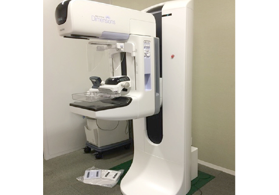
マンモグラフィ
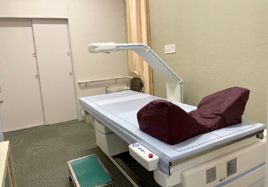
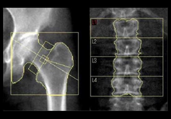
骨塩定量測定
X線を利用して骨密度測定する検査です。当院では、簡易的な測定法ではなく、最も信頼性の高いDEXA法による測定法で検査を行っています。主に腰椎・大腿骨近位部で測定します。測定結果は骨粗しょう症の診断・経過観察や治療判定に用います。この検査は、FAX予約に対応しております。
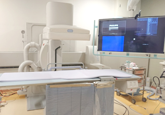
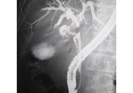
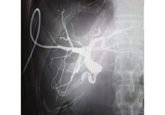
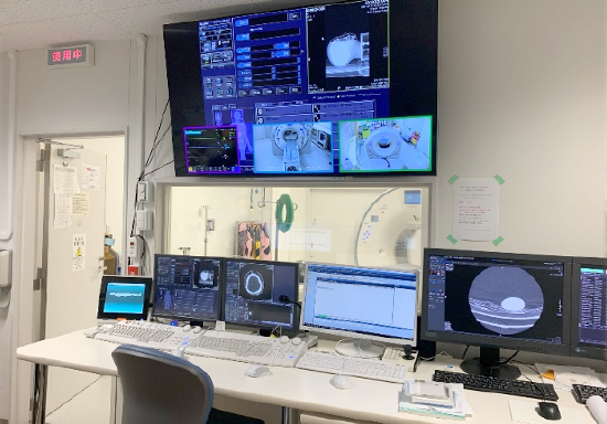
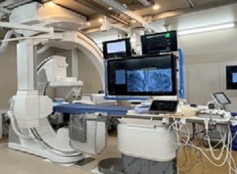
当院のTV・内視鏡部門はCアーム型のデジタルX線TV装置が2台と他3台のX線TV装置が稼働しています。全ての装置にFPD(フラットパネルディテクタ)を搭載したシステムが設置されており、画質の向上と被ばくの低減に取り組んでいます。複数台保有により、検査に対して柔軟に対応することができ、効率的な運用に努めています。また24時間365日緊急検査・治療にも対応可能で2024年度は年間約3900件の検査および治療を行いました。
写真にある大モニタ付きのCアーム型デジタルX線装置では主にERCP、PTCDなど消化器内科による肝・胆・膵領域の検査および治療を行っており、同様のもう1台の装置では、主に呼吸器内科による気管支鏡検査、整形外科による脊髄腔造影検査、さらにほか3台のTV透視装置では、泌尿器科、外科、放射線科、内科全般による検査および治療が行われています。
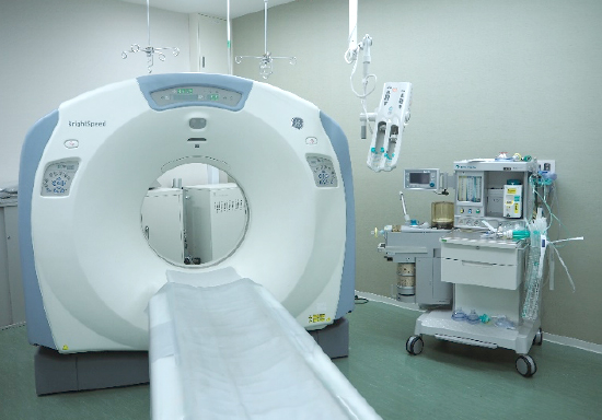
CT検査とは
CT(Computed Tomography:コンピュータ断層撮影)とは、装置の中心に置いた身体にX線を当て、透過したX線の情報をコンピュータで解析することにより、体内を輪切りにした画像を得る撮影方法です。短時間で広範囲の撮影が可能で、撮影したデータからは、様々な方向の断面で切り取ることができ、3D画像を作成することもできます。
また、「造影剤」という薬を静脈から注入して撮影することで、血管や病変などのより詳しい情報を得ることができます。
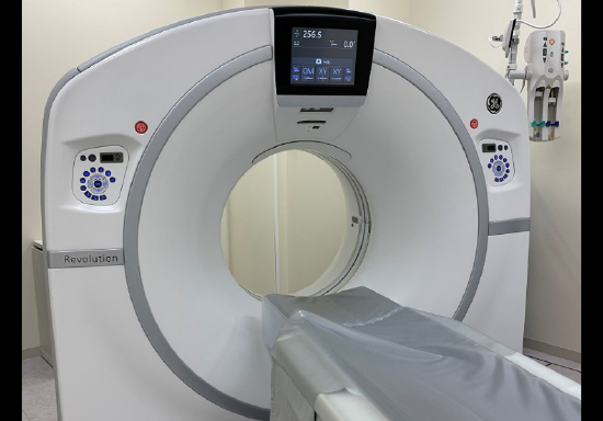
当院のCT装置
| 本館 |
|
|---|---|
| 南館 |
|
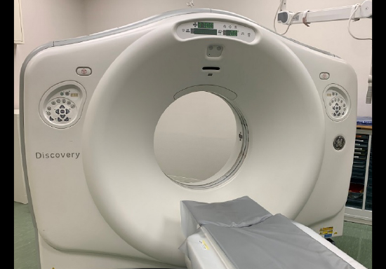
320列ADCTでは、年間約1500件の心臓冠動脈CTを実施し、アブレーション・TAVI・バイパス手術への画像支援を行なっています。
またデュアルエナジーCTでは、通常のCT検査よりも多くの情報を持った画像の提供や、少ない造影剤量での検査を実施しています。
検査に関する注意事項
次項に該当する方は、事前に医師とご相談ください。
CT検査全般について
- 妊娠中、又は妊娠をしている可能性のある方
- 心臓ペースメーカ、埋込み型除細動器(ICD)を使用している方
造影検査をする場合
- 過去にヨード系造影剤に対するアレルギーがあった方
- 気管支喘息治療中の方または喘息薬使用後3年以内の方
- 重篤な心障害、肝障害、腎障害がある方
- ヨード過敏症、甲状腺機能亢進症の方
造影検査を受けられる方へ
造影検査とは造影剤を使用することで組織間のコントラストを向上させ、画像の情報量を増やします。また、血管や腫瘍などを明確に描出することが可能になります。造影剤は腕などの静脈より注入します。注入時に身体が熱く感じられることがありますが、薬の性質によるものなので心配要りません。
造影剤アレルギーについて
- 造影剤アレルギーには、くしゃみや発疹、かゆみ、吐き気からショック症状に至るものまであります。変わったことがあればすぐにお伝えください。また、検査終了してしばらく経ってから症状が出る場合もあるので、その場合には当院までご連絡ください。
造影検査についての注意事項
- 造影剤の検査を受けるためには、検査日より6ヶ月以内の腎臓の機能を診る採血データ(eGFR)が必要になります。これは、造影剤が腎臓を通して排出されるからで、腎臓の機能が著しく低い方には造影剤を使用しない単純検査に変更する場合があります。
- 造影剤は尿に混ざって排泄されます。検査後は、意識して多めに水分を取るようにしてください。
- ビグアナイド系糖尿病薬を服用している方は検査後に約2日間休薬することになります。
- 当院では造影検査後の授乳制限は設けていませんが、ご不安な方は主治医にお申し出ください。
検査の流れ
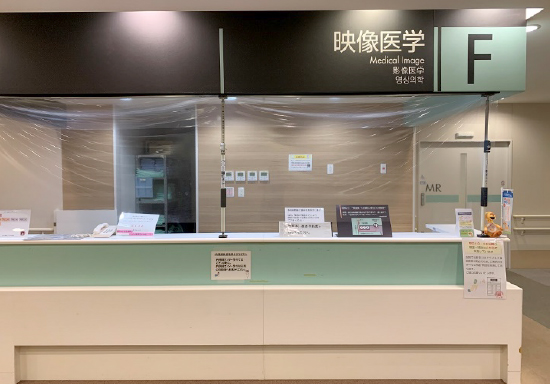
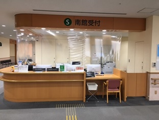
~受付~
| 本館 | 1階「F 映像医学受付」 |
|---|---|
| 南館 | 2階「S 南館受付」 |
注)来院時間の指示を受けている場合は、指示通りの時間にお越しください。
CT検査前に採血データが必要で、採血指示がある場合は予約時間の1時間前までに採血を行ってください。
また、心臓検査の患者様で、検査当日に心拍を抑えるための薬の有無を決定するとの指示があった場合に
は、予約時間の1時間前までにお越しください。
~検査前~
検査の内容によっては、検査着に更衣していただきます。検査着は受付でお渡ししますので、更衣室でお着替えください。撮影する範囲に、金属(入れ歯、ヘアピン、ネックレス、カイロ等)が無いようにしてください。更衣後は中待合室でお待ちいただきます。
造影検査の場合
造影剤を用いた検査の場合、造影剤を注入するために腕などの静脈に注射します。その際、過去の造影検査でのアレルギーの有無や、気管支喘息の有無、糖尿病薬の服用等について問診いたします。また、造影剤の使用量を決めるために体重をお訊きします。
~検査中~
検査時間は約5分~20分です。特殊な検査の場合はこれ以上かかる場合があります。検査中は体を動かさないようにお願いします。また、撮影部位によっては、息止めを行います。検査中は常にモニターで観察していますので、ご安心ください。変わったことがあれば、お渡しするブザーなどで教えてください。
~検査終了後~
造影剤を使用した場合は、造影剤の排泄を促すために水分をいつもより多めにとってください。気になることがありましたら、担当スタッフまでお申し出ください。
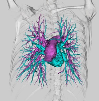
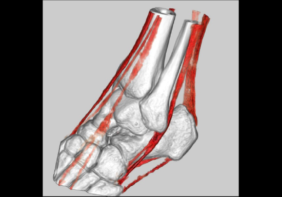
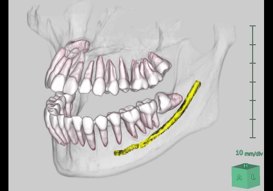
3D-CT
CTのデータから、3D画像を作成することができます。さらに、造影剤を併用することで、疾患と血管や他臓器の関わり合いを視覚的につかむことが容易となり、手術などの治療に有効な画像を提供することができます。
当院ではZioStation2(アミン社製)・VINCENT(富士フイルムメディカル社製)・Advantage Workstation(GE社製)の画像解析装置(workstation)を用いて多くの3D画像を作成しています。
冠動脈3D
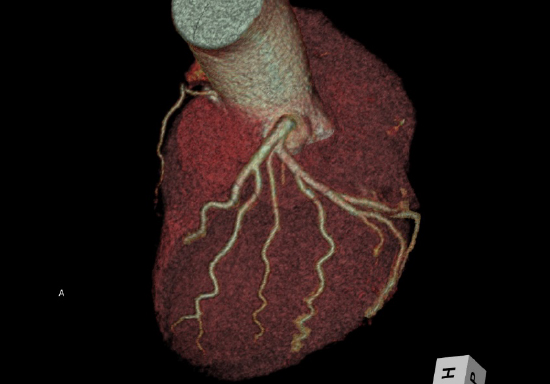
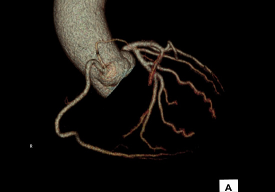
3D-CTA(CT-Angiography)
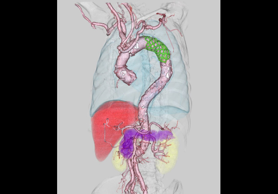
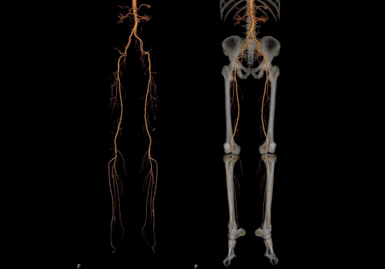
その他3D画像
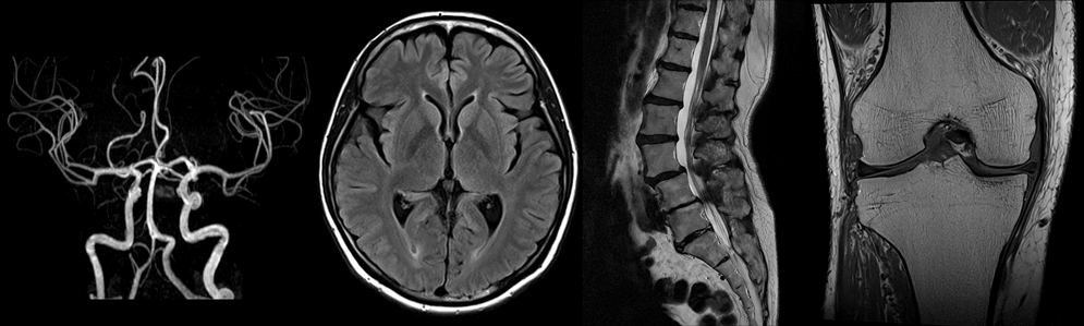
MRI検査とは
MRI検査は強力な磁石でできた筒の中に入り、磁気と電波を使って体の内部を画像化する検査です。
この磁気と電波の影響により、検査中にピリピリと感じたり、体温が上昇することがありますが、検査後の身体に影響を及ぼすことはありませんのでご安心ください。また、放射線を使わずに画像を得るので放射線被ばくの心配はありません。
当院では頭から足先まで全身のあらゆる部位の検査対応を行っています。特に脳、脊髄・脊椎、関節、子宮や前立腺などの診断に有用です。ただし、撮影時間が長いため一度の検査で広範囲の検査対応は行っておりません。
検査には検査内容により約15〜60分ほどお時間を頂いております。
当院のMRI装置
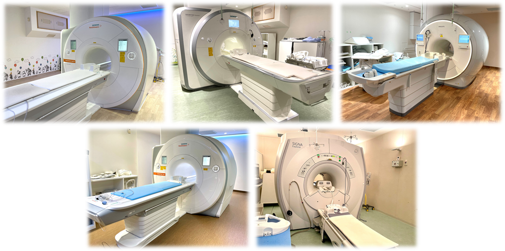
本館1:SIEMENS社製 MAGNETOM Vida fit 3.0T (2024.3~)
本館2:SIEMENS社製 MAGNETOM Avanto fit 1.5T Upgrade (2021.1~)
本館3:Philips社製 Ingenia Elition (2022.2~)
本館4:SIEMENS社製 MAGNETOM Vida 3.0T (2023.6~)
南館:GE社製 Signa Explorer 1.5T SIGNA EXPLORE12 AIR IQ Edition(2021.6~)
注意事項
MRI検査を受けられない可能性のある方
次項に該当する場合、検査を受けられない可能性や検査前後の診察等が必要な場合があります。事前に主治医とご相談下さい。
- 心臓ペースメーカー/人工内耳を埋め込まれている方
- 医療機器(DBS、VNS、SCS、ITB等)を埋め込まれている方
※条件付MRI対応製品であっても、当院の整備・体制から検査を受けられない場合があります。 - 体内に金属を有する方
(人工骨頭・関節、脳動脈クリップ、ステント、コイル、内視鏡クリップ、インシュリン注入ポンプ等) - シャントバルブ挿入中の方
(脳神経外科医師による事前確認が必要となります。当院で対応できないシャントバルブの場合は検査を受けることができません) - 妊娠中もしくは妊娠の可能性のある方
※妊娠13週までの方は検査をお受けいただけません。 - 刺青やアートメイクをされている方
- 閉所恐怖症の方
- 材質が不明な金属が体内にある方
(手術を行った病院や放射線診断科医の事前確認が必要となります。)
また、この他にも安全に検査を受けられないと判断した場合、検査を中断・中止したり、確認のためのお時間を頂いたりする場合がございます。あらかじめご了承ください。
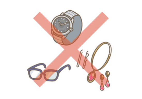
検査室に持ち込めないもの
金属を含む材質のものや磁気を持つものは検査の妨げ、故障の原因や火傷をおこす可能性がありますので、以下のものは取り外してください。
貴金属(時計、指輪、ネックレス、イヤリング、ヘアピン等)
・眼鏡、補聴器、入れ歯、湿布、エレキバン等
・貼り薬、カイロ、ニコチンパッチ等
・グルコース測定器(FreeStyle リブレ等)
・金属のついている下着、保温性素材の下着
(ブラジャー、ヒートテックインナー等)
・カラーコンタクトレンズ、ディファインコンタクトレンズ
・マグネットネイル、ジェルネイル
・磁気カード類(駐車券、診察券、キャッシュカード等)
・金属製のワイヤー入りマスク・金属成分含有のコーティングマスク
(検査前に、全ての方にMRI対応のマスクに交換していただきます。)
※上記以外にも検査室内に持ち込めず外していただくことがありあます。あらかじめご了承ください。
造影検査について
MRI検査においての造影剤の役割は、単純検査(造影剤を使用しない検査)では判別のつかない病気をより詳しく検査するためのものです。次項に該当する場合、造影剤を使用できない可能性があります。事前にお申し出ください。
- 腎機能障害がある方
- 過去にMRIで造影剤を使用し副作用があった方
- 現在喘息の方もしくは喘息の既往がある方/アレルギー体質の方
- 妊娠中の方
- 授乳中の方(ごく微量ですが乳汁中に排泄されることが知られています。安全な量だと考えられますが気になる方は24時間授乳をお控えください。主治医とご相談ください。)
また、心拍を抑えるお薬(β遮断薬)を服用されている方は、造影剤による副作用発生時の対応が異なりますので、事前にお申し出ください。
造影剤は尿から排泄されるため、直近の採血の結果(腎機能の結果)が必要となります。当日に採血の検査がある方は結果を確認するまでにお時間(約1時間)を頂く場合があります。また、造影剤を使用された方は普段より少し多く水分を摂取するようにお願いします。
検査の流れ
~来院前~
安全に検査を受けて頂くために、検査部位に関わらず検査着に着替えて頂きます。当日はできるだけお化粧はせず、貼り薬・アクセサリー類を控えて身軽な服装でご来院ください。また、入れ歯や補聴器、かつら・ウィッグ、ワイヤー付きのマスク等は外して頂きます。
撮影部位(特に腹部や膀胱)によっては、以下のような検査前の飲食・排尿制限がありますので、MRIの検査予約表をご確認いただき、医師の指示に従ってください。
・【絶食3時間】と指示された検査では、必ず検査予約時間の3時間前から絶飲食をお願いします。
・【検査前1時間排尿禁】と指示された検査や医師より指示があった方は、検査予約時間の1時間前までに排尿をお済ませください。
検査にご持参いただくもの
- 診察券
- 予約表
- MRI検査チェックリスト・造影剤使用に対する同意書
~受付~
予約時間の15分前までに受付にお越しください。
| 本館 | 1階「F 映像医学受付」 |
|---|---|
| 南館 | 2階「S 南館受付」 |
チェックリスト・同意書の確認と更衣のご案内をいたします。
~検査前~
更衣室で身に着けている金属や磁気類がないようにしてください。
※詳細は「検査室に持ち込めないもの」をご参照ください。
更衣が終わりましたら、ロッカーに荷物を入れていただき、チェックリスト・同意書を持って待合でお待ちください。
妊娠中や閉所恐怖症など何か気になる点がありましたら、担当技師や看護師にご相談ください。
~検査中~
撮影の際は工事現場のような大きな音がします。ヘッドフォンや耳栓をお渡しします。検査中は動かないようにお願いします。撮影部位によっては、息止めや造影剤を使用します。
※金属製のワイヤー入りマスク・金属成分含有のコーティングマスクは検査中外していただきます。
検査室に案内する際に、MRI対応マスクへ付け替えていただきます。
~検査後~
検査後は更衣していただき、終了となります。
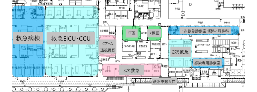
当院にはER型の救命救急センターが設置されており、年間平均33,000名の救急患者と10,000件を超える救急車搬入を受入れており、厚生労働省から発表される「全国救命救急センター評価」においては11年連続全国一位を獲得しています(2025年現在)。
放射線技術部は、救命救急センター内に救急専用のX線室とCT室を備えており24時間365日フル稼働にて対応しています。
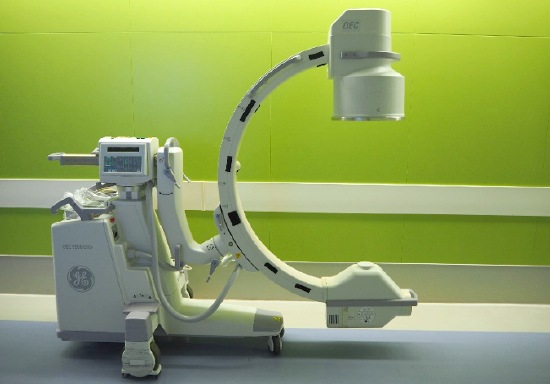
救急専用のFPD搭載ポータブル装置および移動型Cアーム透視撮影装置も備えており、1次救急患者から3次救急患者までのあらゆる救急症例に迅速に対応することができます。
救急外来 装置一覧
| X線撮影装置 | 救急専用X線室 | 島津 | RADspeed pro |
|---|---|---|---|
| FPD搭載 ポータブルX線装置 | 救急病棟 | 島津 | Mobile Art Evolution |
| 移動式Cアーム | 救急外来処置室 | GE | OEC9000 Elite |
| FPD搭載 ポータブルX線装置 | 救急外来 | 島津 | Mobile Art Evolution |
| FPD搭載 ポータブルX線装置 | Covid専用病棟 | 島津 | Mobile Art Evolution |
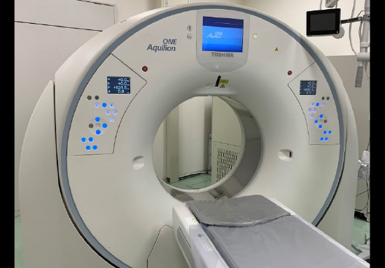
| 320列ADCT装置 | 救急専用CT室 | Canon | Aquilion ONE PRISM Edition |
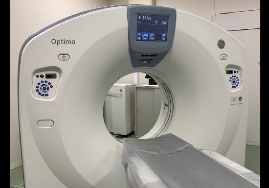
| 64列MDCT装置 | 感染対策CT室 | GE | Optima CT660 |
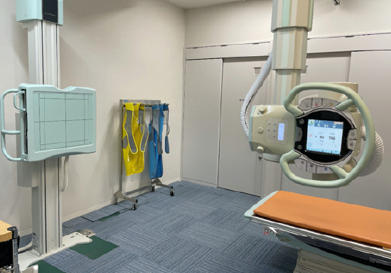
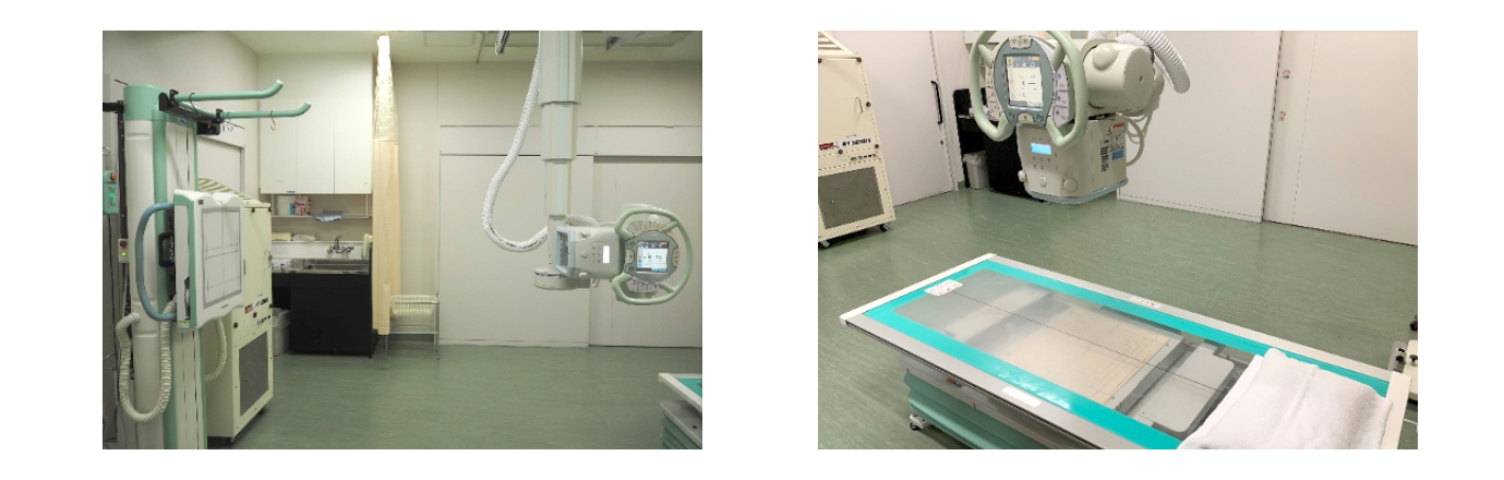
救急専用X線室
救急外来に直結した専用のX線撮影室を設けており、独歩の患者様からベッド搬送の患者様まであらゆる撮影に対応しています。乳幼児撮影に関しても専用の撮影台を有しており、安全に撮影することが出来ます。
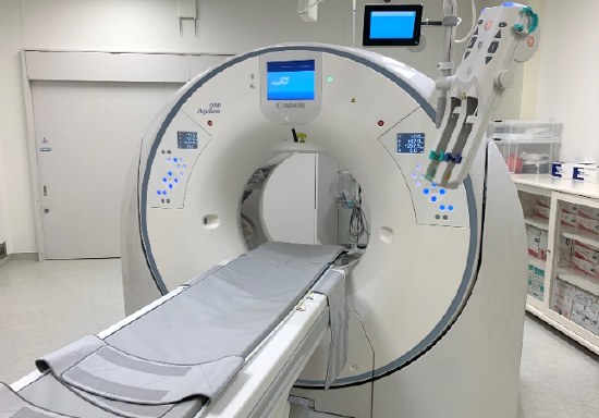
救急専用CT室
2021年に新たに導入された320列ADCT装置は広範囲をブレの少ない画像で提供できる高速撮影、全脳を経時的に撮影することで脳梗塞を高精度に発見できるPerfusion撮影、画像により多くの情報を付加させるDual Energy撮影など最先端の検査が可能です。また、人工知能(AI)を用いて設計された最新の画像再構成技術であるAiCE(Advanced intelligent Clear-IQ Engine)により高品質画像で診断を行えるようになりました。
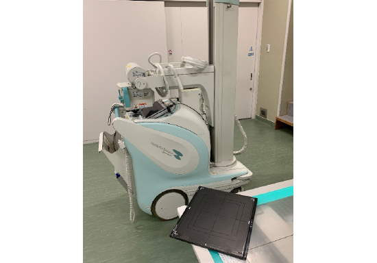
【救急専用FPD搭載ポータブル装置】 【救急専用移動型Cアーム透視撮影装置】
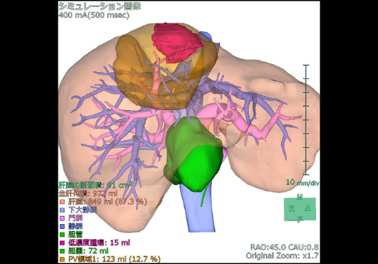
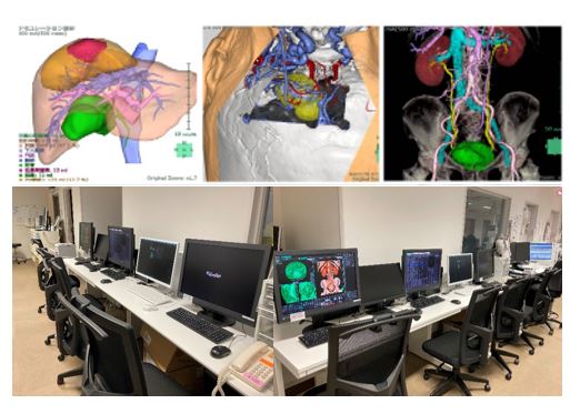
3Dラボ室
2017年より3Dワークステーションを有する画像処理室(3Dラボ)を開設しました。体内の臓器構造を3D化することで手術前に病変と血管、臓器等の位置関係を詳細に把握でき、手術計画の立案や手術シミュレーションなどに活用されています。
CT、MRI、RIなどマルチモダリティで撮影した画像を合成させることで、解剖学的情報と生理学的情報を併せ持った画像が作成でき、臓器の切除範囲などをより精密に決定することが可能になります。また、筋肉量や臓器/腫瘍の容量、血管径などを定量的に測定でき、診断基準の判定材料や定期フォローの客観的な指標として利用できます。3Dラボ室に画像処理専任の技師を配置することで、オペレータの技術の向上と効率化が得られ、従来と比べ質の高い画像処理が可能となり、患者様への低侵襲医療、高度先端医療の提供に貢献しています。
また、院内の電子カルテ端末にネットワーク型の3Dワークステーションを装備することにより、3Dラボ室のみに留まらず院内のあらゆる場所で画像処理が可能であり、医師の要望に迅速に応えています。
3Dラボ室 装置一覧
| 3D画像解析システム | 電子カルテ | 富士フイルムメディカル | VINCENT network |
|---|---|---|---|
| 3D画像解析システム 3台 | 3Dラボ室 | ziosoft | Ziostation2 |
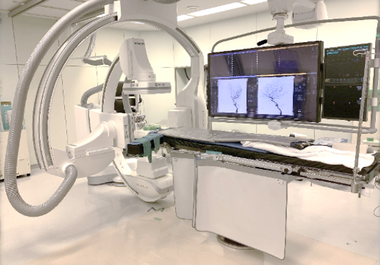
〈第2技術部〉血管造影・手術室・Hybrid
また、血管造影部門は救急外来から1分以内に移動できる場所に位置しており、緊急IVRが必要な救急患者に対する迅速な対応が24時間いつでも可能な体制となっています。
放射線技術部では、チーム医療の一員として迅速な治療を行える環境を整えると共に、「安全で確実な血管内治療を支援する画像の提供」を目指し、日々業務に臨んでいます。
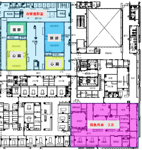
血管造影室 装置一覧
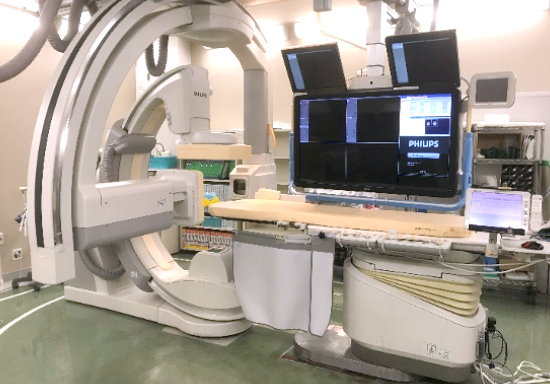
| 頭頸部血管用装置 | 血管造影室1 | PHILIPS | Allura Clarity FD20/15 |
|---|---|---|---|
| 血管造影室2 | Azurion7 B20/15 | ||
| 心臓用血管撮影装置 | 血管造影室3 | SIEMENS | Artis Zee BC PURE |
| 血管造影室4 | PHILIPS | Azurion7 B20 | |
| 血管造影室5 | SIEMENS | Artis Zee BC Polydoros | |
| 腹部血管用IVR-CT | 血管造影室6 | Canon | XTP 8100XG TSX 310C |
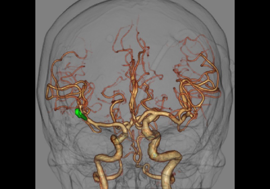
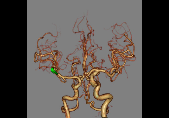
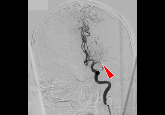
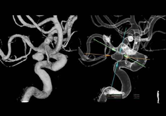
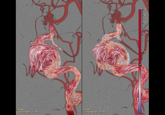
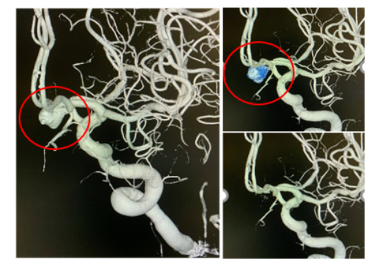
頭頸部領域
頭頸部領域では、「脳動脈瘤に対する血管塞栓術(コイルエンボリ)」「脳梗塞に対する血栓回収術」「頚動脈狭窄に対するステント拡張術」などを実施しています。放射線技術部は機器精度管理に加え、3D画像作成や病変サイズの計測をすることで、医師の診断、治療を支援しています。
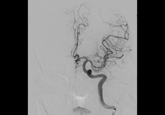
脳卒中に対しては、24時間救急隊員から直接call可能な脳卒中Hotlineを開設しており、脳神経外科・脳神経内科の共同チームで対応しています。特に脳梗塞に対する治療では、1秒でも速く血管を再開通させることが極めて重要です。当院では脳卒中の診断から治療までをタイムロスなく行えるシステムを、医師、看護師、放射線技師がチーム一丸となり構築しています。
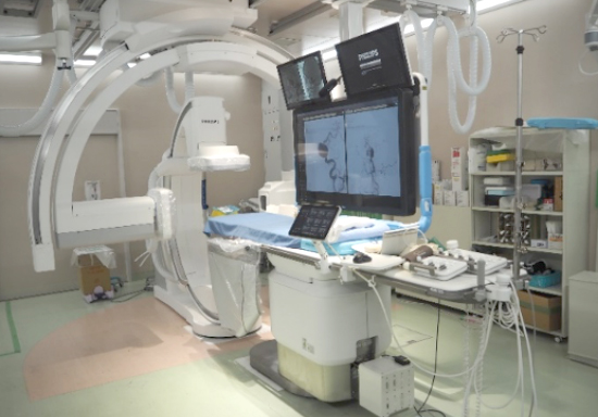
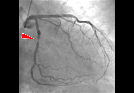
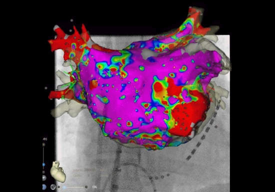
心臓循環器領域
心臓循環器領域ではFPD搭載のバイプレーン血管撮影装置3台を有しており、「虚血性心疾患(狭心症、心筋梗塞など)に対するカテーテル治療」「徐脈性不整脈に対するペースメーカー挿入」などを実施しています。さらに、頻脈性不整脈の代表的な治療方法である「心筋焼灼術カテーテルアブレーション」も積極的に行われており、最先端の3次元ナビゲーションシステムを併用することで、被ばく線量低減を実現しながら、より安全確実な治療が可能となっています。
また、当院では心肺停止患者に対してECPRを導入しています。ECPRとは心臓マッサージをしながら血管造影室に搬送、人工心肺装置(PCPS)の挿入までを行うことで、循環動態を維持した後に、心臓カテーテルによる原因精査と治療を行う手技であり、豊富な経験を持つ医師、高度医療機器を備える設備、そして何より各医療スタッフの息の合った連携が必要不可欠です。ECPRに対するシミュレーショントレーニングを日々実践し、1人でも多くの心肺停止患者の社会復帰を支援できるよう血管造影部門は常に備えています。
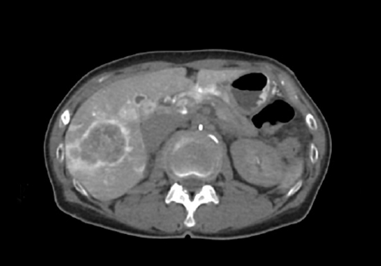
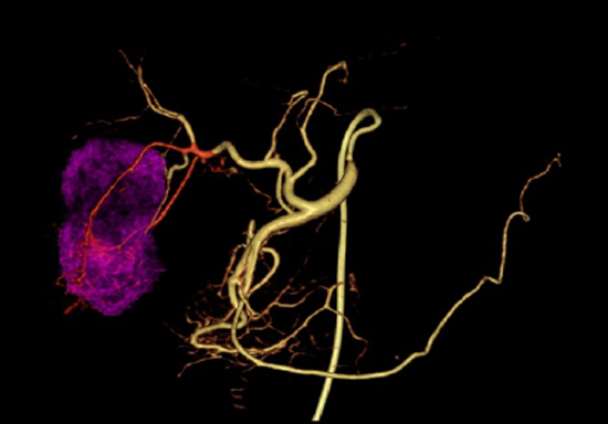
腹部血管領域・その他全身血管領域
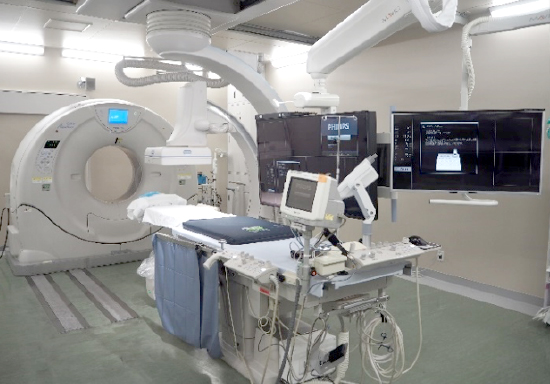
腹部血管領域では、血管撮影装置とCT装置を同室に設置した「IVR-CTシステム」を導入しています。CT装置には320列ADCTを装備しており、形態・動態・機能診断に必要なデータを広範囲に短時間で収集することができます。このIVR-CT 装置では、主に腹部⾎管に対するIVR を実施しています。治療中にCT 画像が撮影できることで、3次元的な⾎管の形態把握が可能になり、カテーテルの適切な経路選択をサポートできます。さらに治療部位の評価や合併症の検索等を⽬的としたCT 検査も、検査室を移動することなく撮影することが可能です。その他にも⾮⾎管系IVR として、CT ガイド下⽣検およびCTガイド下ドレナージなども実施しており、この装置を使⽤することで、安全で効率的に⼿技をサポートすることが可能となりました。
放射線技術部は機器操作を行い、治療中に撮影した画像作成・計測を行うなど医師の支援をしています。

手術室部門では、手術室・ICU専用のFPD搭載ポータブル2台と、移動式Cアーム4台を導入し、19室あるオペ室とICUの撮影に対応しています。また、手術室とICUに隣接する位置にCT装置を設置することで、手術直後の患者様や、ICU入院中で生命維持装置使用中の患者様など、緊急性が高く移動にリスクがある患者様の診断を迅速かつ安全に行えるようにしています。
また、手術室部門には「ハイブリッド手術室システム」を2室装備しています。ハイブリッド手術室とは、手術室と血管透視撮影装置を組み合わせたシステムのことで、従来血管造影室で施行していた血管内治療が、手術室でより安全に行えると共に、手術室と血管造影室、それぞれ別の場所に設置されていた機器を組み合わせることにより、最先端の医療技術に対応しています。
手術室・ICU 装置一覧
| 移動式Cアーム 9inch 2台 | 手術室 | GE | OEC9000 Elite |
|---|---|---|---|
| 移動式Cアーム 12inch 2台 | 手術室 | GE | OEC9000 Elite |
| FPD搭載 ポータブルX線装置 2台 | 手術室 ICU |
日立 | シリウス HP130 |
| CT装置 | 手術室 ICU |
GE | Bright Speed Elite |
| ハイブリット シングルプレーン | 手術室1 | PHILIPS | Azurion7 FrexArm |
| ハイブリット バイプレーン | 手術室4 | SIEMENS | Artis Q BA Twin |
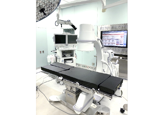
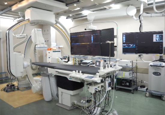
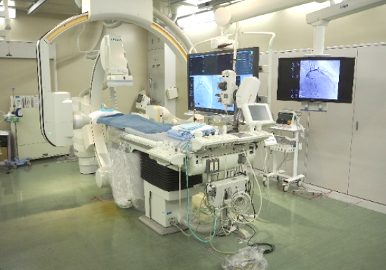
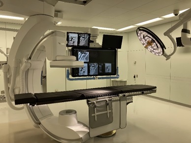
手術室1 ハイブリッド・シングルプレーン
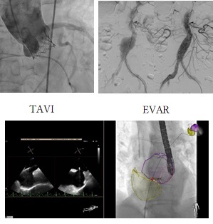
手術室1は循環器領域の手術に利用されており、2022年4月に装置が更新されて最新鋭のハイブリッド血管撮影装置となりました。本装置の特徴として、高画質低被ばく、自由に移動可能なCアーム、また、透視像と超音波装置の融合画像も可能です。さらに映像モニターが充実して映像表示も自由自在に変更することが可能です。手術としては、経カテーテル大動脈弁留置術(TAVI)、大動脈ステントグラフト留置術(TEVER・EVAR)、ASD、PFO、ウォッチマン、ペースメーカー留置等様々な手術に使用されています。
手術室 ハイブリッド・バイプレーン
手術室4は主に脳神経外科領域の手術に使用しており、バイプレーン透視撮影装置を設置しています。手術中の透視装置の併用により、造影剤を使用した3D画像の構築が可能なため詳細な血管評価ができ、治療成績と安全性の向上が図れます。
また、脳動脈瘤に対する開頭クリッピング後の評価などに使用され、開頭部分を閉じる前に確実な手術効果判定が可能となり、再手術のリスクを抑えることができます。
手術室4
〈第3技術部〉放射線治療・核医学・PET
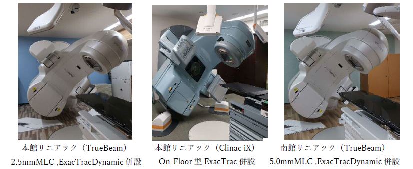
外部照射
外部照射は、リニアック(直線加速器)を用いて高エネルギーのX線や電子線を体の外から照射して治療を行います。当院では3台のリニアックを有しており、画像誘導放射線治療(IGRT)可能な装置で、高精度な多門照射・強度変調放射線治療(IMRT)・定位放射線治療(SRT)が実施可能です。症例により呼吸同期システムであるRGSC(Respiratory Gating for Scanners)を使用することにより呼吸性移動を考慮した治療も可能です。
2.5mm幅のビーム絞り機構(マルチリーフコリメータ:MLC)を搭載したTrueBeamは、脳転移などの小さい病変や複雑な形状の病変に対して、これまで以上にフィットした照射野を作ることが可能となり、正常臓器への線量低減が可能です。また、フラットニングフィルタフリー(flattering filter free:FFF)での高線量率X線照射が選択可能であり、IMRTやSRTを短時間に行うことが可能であり、息止め照射時の患者負担も減らせます。
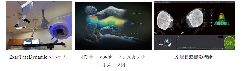
ExacTracシステム
リニアックがある部屋の床と天井にX線撮像装置を設置し、そのX線の画像情報を元に患者の位置照合を6軸補正で行い、高精度放射線治療を実現するシステムです。
ExacTracDynamicシステムは、新技術となる4Dサーマルサーフェスカメラを導入し、従来のX線画像誘導と熱分布による体表面誘導を組み合わせることで治療中の患者位置の照合と補正を行うことが可能です。
また、X線の自動撮影機能により、リニアックのビーム照射中のX線画像照合が可能で、任意のガントリ角度においてX線画像照合を行い、閾値を超えた誤差を検出した場合は、リニアックの照射を中断することが可能となりました。
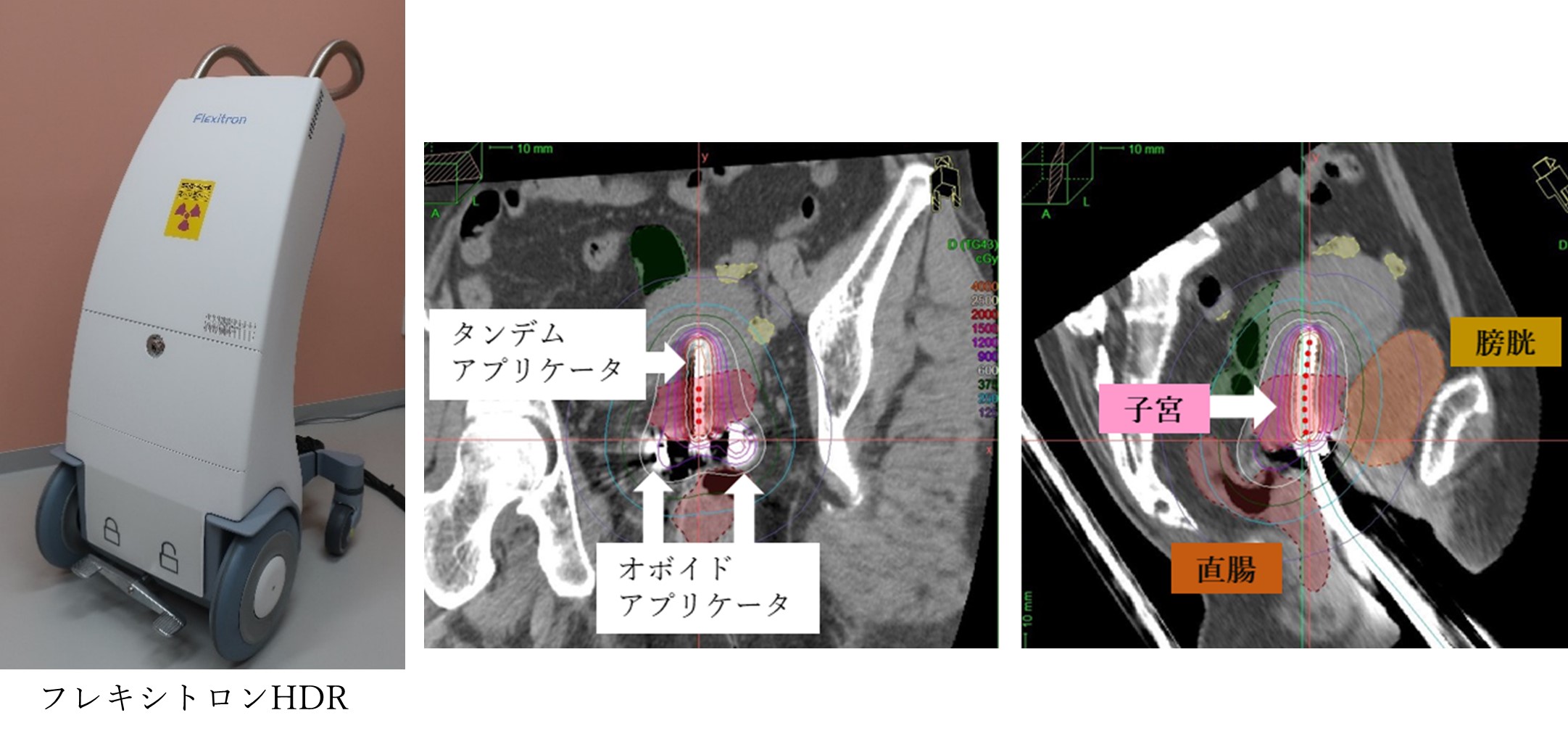
高線量率密封小線源治療装置(Remote After Loading System:RALS)
密封小線源治療は、放射線の線源を病巣の近くや内部に挿入し、体の内側から放射線を照射する治療法です。線源に近いほど線量は高く、線源から距離が離れると急激に線量が減少するため病巣には高い線量を、周囲の正常組織には線量を低く抑えることができます。
当院では、婦人科癌(子宮頸がん・子宮体がん・膣がん)を主な対象疾患としており、外照射と組み合わせて治療を実施しています。1回の治療に要する時間は、治療準備~治療計画~照射までの一連で約3時間、週1回の治療を3~4回行います。2024年5月にフレキシトロンHDRに装置を更新し、より精密な治療を行えるようになりました。現在は、組織内照射併用腔内照射の臨床開始に向けて、準備を進めています。
永久刺入組織内照射(125I)
前立腺癌に対する密封小線源治療は、「シード線源(125I小線源)による前立腺永久挿入療法」を実施しています。
放射線治療について詳しく知りたい方は放射線治療科へPET/CT
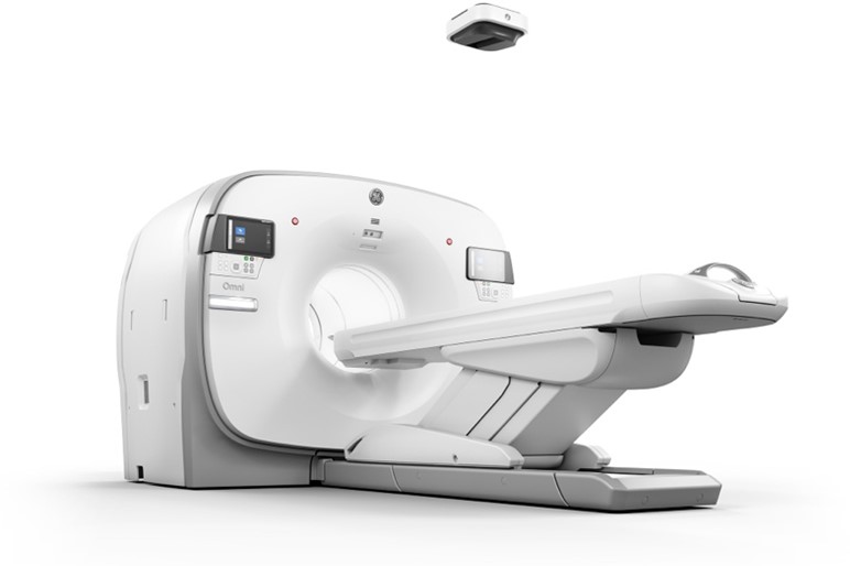
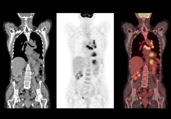
呼吸同期が可能な精度の高い融合画像が得られ、明確に病変部位や範囲が診断でき、全身の腫瘍検索に優れているPET/CTを3台導入しています。FDG-PET検査、アミロイドPET検査を実施しています。これらの検査は他院からの依頼も受けております。今年度より脳腫瘍に対するPET検査を開始いたします。また、半導体を搭載した最新のPET/CT装置が導入されます。
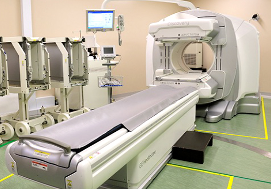
SPECT/CT
一度の検査で解剖学的位置情報と機能情報が得られ、吸収補正による画質向上を可能としたSPECT/CT 1台に加え、16列のMDCTを搭載したSPECT/CT 1台を導入し診断精度の高い検査を提供しています。
主に骨シンチ、脳血流シンチ、心筋血流シンチ検査を実施しています。
放射性同位元素内用療法
177Luを用いた神経内分泌腫瘍に対する内用療法、131Iを用いた甲状腺がん転移巣への内用療法を糖尿病・内分泌内科と協力して行っています。これまで入院のみで行っていた131Iを用いた内用療法でしたが、病状によっては外来でも内用療法を施行することができるようになりました。放射線治療科と協力して、223Raを用いた去勢抵抗性前立腺がん骨転移巣への内用療法を実施しております。
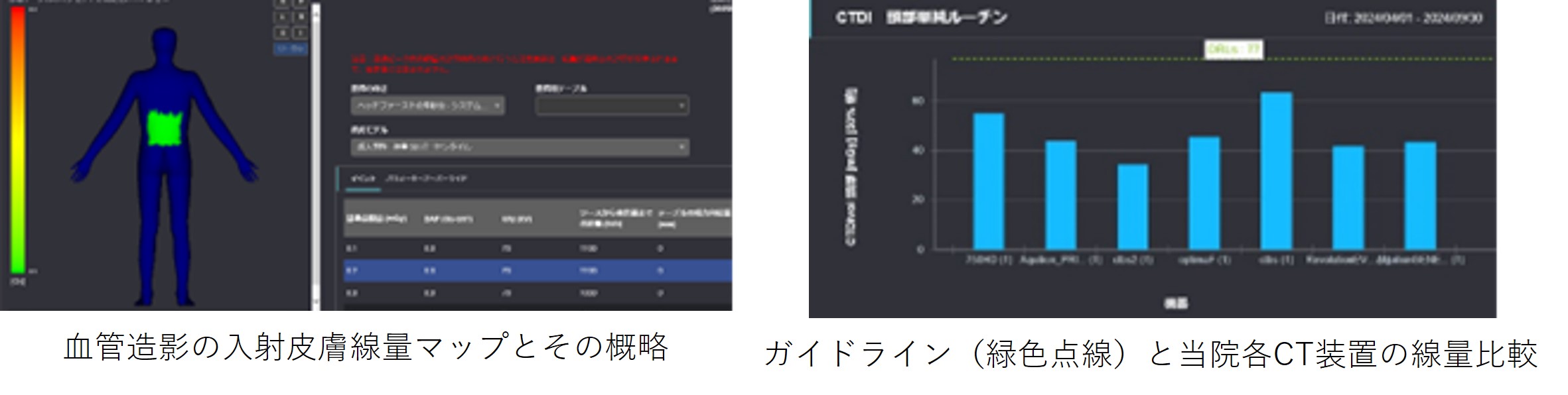
医療放射線の適正な管理について
放射線を用いた検査(放射線検査)は、患者様の診断や治療に大きく役立てられています。その一方で、これらの検査では放射線による被ばく(医療被ばく)は避けられません。
当院では令和4年度にバイエル薬品社製の「Radimetrics」(医療放射線の情報を一元的に管理できるシステム)が導入されました。このRadimetricsを使用することでCT検査、血管造影及び核医学検査等において用いられた放射線量の詳細なモニタリングが可能となりました。
そして、Radimetricsから得られた線量情報を用いて、診療放射線技師は医療被ばく線量に対して検査の目的や画質などのバランスを考慮したうえで、各検査において被ばくの低減に努めるとともに、日々最適な条件で放射線検査が実施されるよう装置の管理を行っています。
もし、放射線検査に関してご不明点やご不安なことがございましたら、主治医もしくは検査スタッフにお尋ねください。
地域医療機関の先生方へ・業務実績
現在、FAX予約で紹介をお受けできる検査はCT検査、MR検査、骨塩定量測定が中心となっております。CT検査はすべて、撮影時間が短く高精度なMDCTで対応しますので、息を止めるのが困難な高齢の患者様にも大変効果的です。また28年3月より冠状動脈CTには、最新320列CT装置を導入しました。革新的被ばく低減機能と超高速撮影により、長時間の息止が不要で不整脈のある患者様にも、より低被ばくで高鮮鋭度の画像が構築できるようになりました。高精細画像により、微小病変などの診断能が確実に向上することが期待できますので、他のモダリティーも含めましてもっと手軽に当院の画像診断装置を利用していただき、患者サービスの向上と地域連携の強化を図っていきたいと考えております。
地域の先生方におかれましても、今まで以上にFAX予約を活用していただき、診療のご支援となりますよう一層のご利用をお願い申しあげます。
放射線技術部の主な業務量
| 2020年度 | 2021年度 | 2022年度 | 2023年度 | 2024年度 | ||
|---|---|---|---|---|---|---|
| CT | 総件数 | 43,152 | 47,497 | 51,343 | 52,406 | 54,332 |
| 時間外件数 | 7,522 | 8,650 | 10,393 | 10,623 | 11,102 | |
| MR | 総件数 | 18,131 | 19,413 | 19,243 | 20,658 | 20,536 |
| 時間外件数 | 948 | 1,144 | 1,178 | 1,206 | 1,071 | |
| 一般撮影 | 総件数 | 52,393 | 56,479 | 58,606 | 62,616 | 68,486 |
| 時間外件数 | 4,614 | 5,013 | 5,390 | 6,421 | 7,568 | |
| ポータブル撮影 | 総件数 | 23,932 | 29,090 | 29,726 | 23,144 | 25,888 |
| 時間外件数 | 8,184 | 11,328 | 11,225 | 9,328 | 10,862 | |
| 特殊撮影 | 乳房撮影 | 1,075 | 1,189 | 1,480 | 1,826 | 1,849 |
| 歯科パノラマ等 | 2,425 | 3,049 | 2,844 | 2,674 | 2,746 | |
| 骨塩定量 | 1,117 | 1,281 | 1,589 | 1,817 | 1,890 | |
| X線TV・内視鏡 | 総件数 | 3,666 | 3,421 | 3,957 | 3,661 | 3,848 |
| 血管造影 | 頭頚部系血管 | 990 | 1,111 | 1,084 | 973 | 802 |
| IVR | 290 | 295 | 367 | 225 | 180 | |
| 循環器系血管 | 1,604 | 1,741 | 1773 | 1,710 | 1,695 | |
| IVR | 1,051 | 1,115 | 1,253 | 1,049 | 1,092 | |
| 腹部四肢血管 | 494 | 747 | 625 | 633 | 611 | |
| IVR | 318 | 449 | 427 | 394 | 377 | |
| ハイブリッド 手術室 |
総件数 | 200 | 233 | 251 | 356 | 396 |
| TAVI | 57 | 86 | 101 | 86 | 102 | |
| ステントグラフト | 32 | 28 | 42 | 40 | 47 | |
| 核医学検査 | SPECT・その他 | 2,014 | 2,141 | 2,099 | 1,943 | 1,932 |
| PET | 2,752 | 2,725 | 2,746 | 2,710 | 2,870 | |
| 放射線治療 | 新患者数 | 656 | 719 | 685 | 643 | 708 |
| IMRT新患者数 | 88 | 98 | 61 | 76 | 114 | |
| 定位治療新患者数 | 65 | 83 | 79 | 61 | 91 | |
| TBI新患者数 | 20 | 22 | 22 | 21 | 26 | |
| 体腔治療新患者数 | 16 | 21 | 26 | 12 | 19 | |
臨床研究・学術実績
| 研究課題名 | 説明文 (PDF) |
|---|---|
| PET/CTにおける呼吸同期技術(Data-Driven Respiratory gating)に関する検討 | |
| ドパミントランスポーターシンチグラフィ検査における解剖学的標準化が定量解析に与える影響の検討 | |
| 線量管理システムを用いた核医学検査の実効線量調査 | |
| 腹臥位心臓CTでの左心耳造影効果に関する造影剤注入方法の検討 | |
| 骨シンチグラフィにおける正常骨SUVの検討 |
レジデント制度について
概要
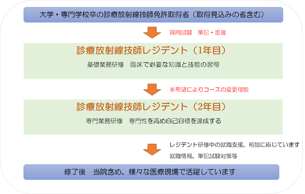
当院の診療放射線技師レジデント制度は、実務経験に基づいた講義と臨床実務実習を通じて、高度急性期医療に対応した診療放射線業務並びにチーム医療を実践できる診療放射線技師の養成を目指します。
レジデントは希望する専門性に合わせて、画像診断コース、放射線治療コースいずれかを選択し、各コースのカリキュラムに沿って研修を行います。
各コースで内容に違いがありますが、一年次には臨床実務経験を積むことを主眼とし、臨床で必要な知識と技能の習得を目指します。
二年次には自己目標や専門性に合わせて、専門業務研修を行い、専門性を高め、自己目標の達成を目指します。また部門のカンファレンスに参加し、医師や他職種と積極的に関わり、質の高いチーム医療と他職種連携を学びます。
目標
実務経験に基づいた講義と臨床実務実習を通じて、高度急性期医療に対応した診療放射線業務並びにチーム医療を実践できる診療放射線技師の育成を目標とする。
目的
画像診断コース
一般撮影、CT、MR、血管造影、救急業務、核医学等についてOJTを中心とした広範な臨床実務経験を積むことを目的とする。
放射線治療コース
基礎的画像診断の研修を行い、放射線治療部門にて、標準測定法ならびに当院で実施する放射線治療分野の実務経験を積むことを目的とする。
年間スケジュール
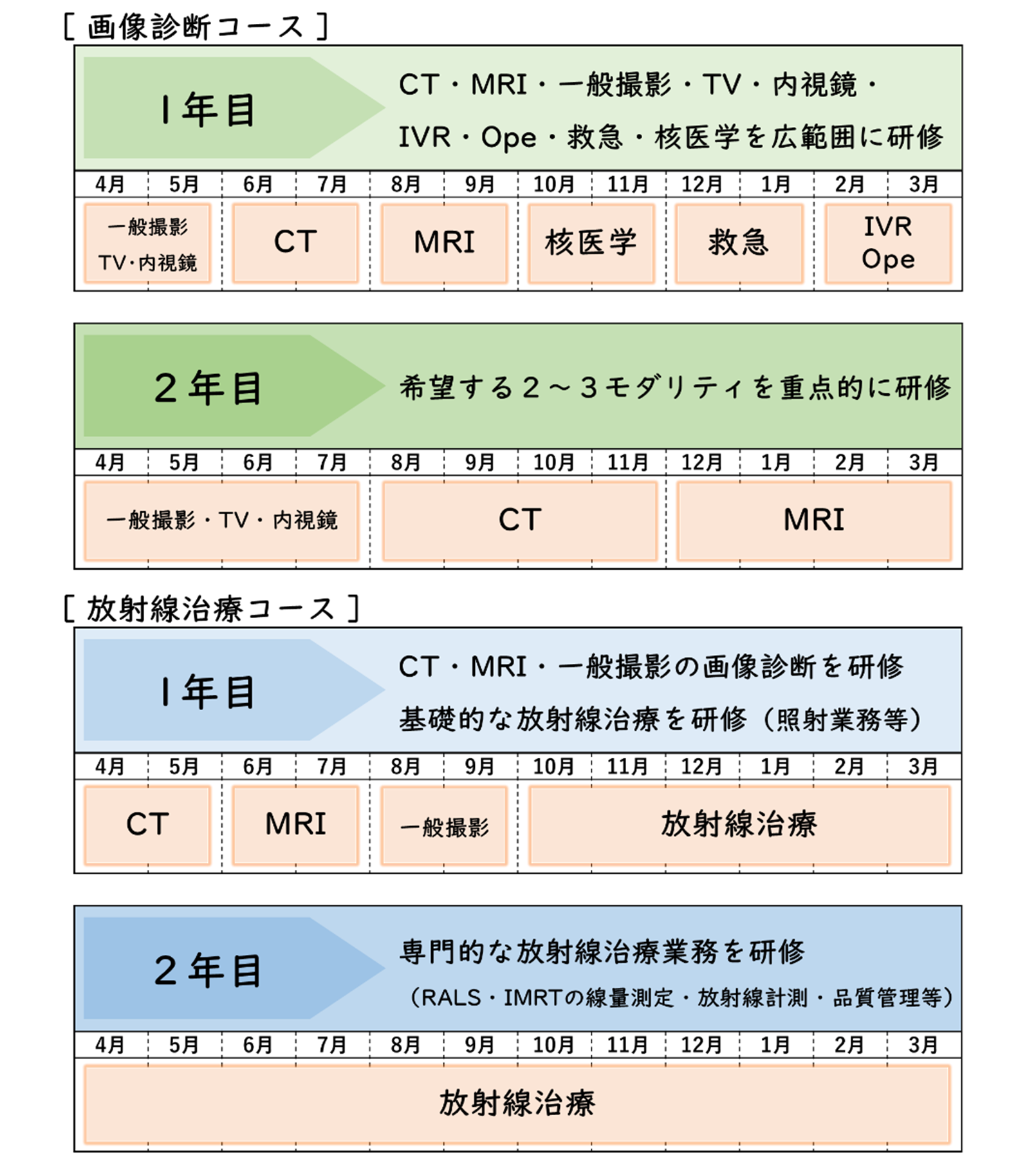

※上記は一例です
※各モダリティの研修時期がレジデント同士で重ならないように研修を行います
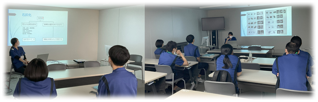
講義研修
毎月、部内でレジデント育成チームによる研修会を開催しています。研修会の前半には、研究発表の能力を育成するため、年3回のレジデントによる症例報告の場を設け、後半には、レジデント育成チーム担当者による講義や症例検討会を行っています。
| 4月 | 5月 | 6月 | 7月 | 8月 | 9月 | |||
|---|---|---|---|---|---|---|---|---|
| 研修内容 | 前半 | 題目 | オリエンテーション | 症例報告 | 症例報告 | 症例報告 | 講義 | 症例報告 |
| 発表者 | 1年目レジデント | 2年目レジデント | 1年目レジデント | 2年目レジデント | 1年目レジデント | |||
| 後半 | 題目 | 感染管理 | 医療安全 | 造影剤 | 医療情報 | 装置管理 | ||
| 発表者 | レジデント育成チーム担当者 | |||||||
| 10月 | 11月 | 12月 | 1月 | 2月 | 3月 | |||
|---|---|---|---|---|---|---|---|---|
| 研修内容 | 前半 | 題目 | 症例報告 | 症例報告 | 講義 | 症例報告 | 症例報告 | 1年間のまとめ |
| 発表者 | 2年目レジデント | 1年目レジデント | 2年目レジデント | 1年目レジデント | 2年目レジデント | |||
| 後半 | 題目 | 画像処理 | 患者移乗 | 患者接遇 | インシデントレポート | 放射線治療 当院の概要 |
||
| 発表者 | レジデント育成チーム担当者 | |||||||
主な就職先
- 地方独立行政法人 神戸市民病院機構
- 神戸大学医学部付属病院
- 滋賀医科大学医学部付属病院
- 公益財団法人附興風会 医学研究所北野病院
- 三菱神戸病院
- 独立行政法人 国立病院機構
- 地方独立行政法人 市立吹田市民病院
先輩の声
Q&A
A.福利厚生について違いはありません。正規職員と同様にベネフィットステーションというサービスが利用可能です。職務内容についてはレジデントに雑務はなく、研修プログラムに従って、研修に専念できます。
A.休日勤務や当直などはありません。
A.4月1日から勤務した場合、10月1日に有給休暇が10日間付与されます。
採用情報
業務内容
放射線技術部は、診療放射線技師・医学物理士、合わせて60名を超えるスタッフが在籍しています。高度医療機器を活用し、先進的な画像診断・治療・IVR等の支援画像の提供、さらに治験・臨床研究分野にも携わっています。また国内有数の救命救急センターを擁する当院は、「神戸市民最後の砦」としての使命から、MRI検査・IVR(血管造影室・ハイブリット手術室等)等の高度医療を24時間体制で行っています。医療は日進月歩です。医療安全・チーム医療・患者サービス向上を基本に医療人としての責任感と協調性を持ち、現状に満足せず、知識・技術力向上に向けて研鑽して頂きたいと思います。専門技師資格を有するスタッフも多数在籍しています。
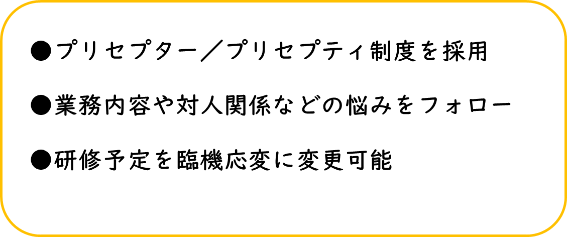
教育体制
入職1年目~3年目まではプリセプター制度を実施しています。1年目として社会人としての姿勢や基本スキルの習得等から始まり、判断力・コミュニケーション能力等の医療職としての基礎を身に着けた人材を育成して参ります。自己研鑽が基本ですが、できる限りのサポートは行って参りますので安心して下さい!又、放射線技術部内での定期的な研修会も実施しています。なお、学会・院外の研修会の参加・専門技師取得に関して一部支援制度を設けていますので、キャリアアップ・スキルアップに活用頂ければと思います。
はたらく環境について
放射線技術部は比較的年齢層が若いので気軽に質問ができ、上司・先輩においても意見交換がしやすい環境であると思います。「育児・介護休業法」に基づく男性技師の育児休業も取得されています。神戸は東西南北が分かりやすく、北は六甲山・有馬温泉、南はハーバーランド・メリケンパーク、中心地には異人館・南京町、神戸ビーフ・神戸スイーツ等の多くの観光スポット・グルメがあります。現在はコロナ禍の為、自粛せざるを得ませんが、機会があれば足を運んでみてはいかがでしょうか?きっと良いリフレッシュになると思います。
先輩からのメッセージ
プリセプター制度により、一対一の関係で先輩から指導していただけます。クリニカルラダーに基づいた目標達成のプランを一緒に考え、気にかけてくださるので自身の進歩状況を確認しながら働くことができます。仕事内容で困ったことがあった時でも質問しやすく、安心できる職場環境です。さらに、社会人として働く上での考え方などを身近な先輩から学ぶことができるため、精神的な面でも自身の成長につながります。ただ撮影技術を学ぶこと以上の成長ができるこの病院でぜひ一緒に働きましょう!(女性技師:入職1年目)
入職して3年目となります。1年目の当直研修後、部署ローテーションで血管造影、一般撮影を経て、現在はCT部門に所属しています。2年目からは月2,3回の当直業務にも従事しています。
3年目では初期研修プログラムの一環で、プリセプティーとして先輩に指導してもらうだけでなく、1年目のプリセプターも担当しました。教えながら自分の知識不足など改めて気付くことも多く、コミュニケーションの取り方や人材育成など、自分自身の新しい勉強の機会にもなりました。
日々の業務に追われることもありますが、年齢の近い先輩も多く、丁寧に教えてくださり、困ったことも気軽に相談しやすい環境です。放射線技師として大きく成長できる職場だと感じています。(女性技師:入職3年目)